Revolutionary Imaging Method Unveils Hidden Brain Structures for Advanced Tumor Diagnosis
February 1, 2024A groundbreaking microscopy technique, developed by MIT and Brigham and Women’s Hospital/Harvard Medical School researchers, is set to transform brain tumor diagnosis and treatment. This novel imaging approach, offering unprecedented high-resolution insights, delves into human brain tissue at the nanoscale, uncovering previously invisible cells and structures.
Key Highlights:
- Unprecedented Detail: The new microscopy technique surpasses previous imaging capabilities, providing researchers with an unparalleled view of human brain tissue. This breakthrough allows for the identification of intricate structures and cells that were once beyond the scope of visibility.
- Revelations in Tumor Aggressiveness: In analyzing brain tumor samples, the researchers discovered unexpected levels of putative aggressive tumor cells in so-called “low-grade” tumors. This surprising finding suggests that certain tumors may possess a higher degree of aggressiveness than conventionally believed.
- Clinical Implications: The imaging method holds the potential to revolutionize tumor diagnostics, prognosis accuracy, and treatment selection. By enhancing the understanding of brain tumor growth and progression, this tool could become instrumental in guiding personalized treatment strategies.
Expansion Microscopy Technique:
The imaging technique is rooted in expansion microscopy, a method developed in 2015. Unlike conventional high-resolution imaging with specialized and expensive microscopes, expansion microscopy involves enlarging the tissue itself. By embedding tissue into a swelling polymer and breaking apart tissue-holding proteins, the researchers achieve a 70-nanometer resolution, previously attainable only with advanced microscopes.
Decrowding Human Brain Tissue:
Overcoming challenges in working with human brain tissue, the researchers developed a tissue-softening protocol to break down chemical treatments while preserving proteins. This allowed the labeling of up to 16 different molecules per tissue sample, including markers for structures like axons and synapses, cell types, and molecules linked to tumor aggressiveness and neurodegeneration.
The imaging technique was applied to brain tumor samples, distinguishing between high-grade glioblastoma and low-grade gliomas. The unexpected abundance of aggressive tumor cells in low-grade gliomas highlights the potential for this tool to redefine the understanding of tumor biology at the nanoscale.
The researchers envision the expansion microscopy technique as a diagnostic tool capable of identifying marker cells and interactions that were previously inaccessible. As clinicians leverage this simple yet powerful tool, it opens up possibilities for neuro-oncology and neuropathology to explore neurological diseases at the nanoscale, providing a deeper understanding for improved patient outcomes.
In conclusion, this revolutionary imaging method holds promise in reshaping the landscape of brain tumor diagnosis and treatment, ushering in an era of precision medicine with nanoscale insights.
Related posts:
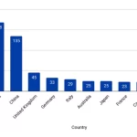
![Country affiliation for papers in all bioinformatics RCs]()
Unveiling the Landscape of Bioinformatics: Insights from Research Clusters
news![Direct RNA Sequencing (DRS)]()
MGI Tech's DNBSEQ-E25 and G99 Platforms Achieve Groundbreaking Sequencing Milestone on Mount Everest
news![metagenomics]()
Scientists Unveil "Obelisks": Mysterious Entities Found in Human Microbiome
news![AI-antibiotic-research]()
Unraveling the Reversibility of Antibiotic Resistance: A Game-Changing Discovery
news![AI based search]()
AI's Impact on Search: Balancing Innovation, Economics, and Ethics
news![will bioinformatics replace by AI]()
Transforming Medical Coding: AI-Driven ICD-10 Solutions
A.I![Maximizing Efficiency: How 5 AI Tools Revolutionized My Workflow]()
Maximizing Efficiency: How 5 AI Tools Revolutionized My Workflow
A.I![Computer-vaccine-design-bioinformatics]()
2025's Breakthrough Trends in Bioinformatics: AI, Genomics, and Personalized Medicine Reshaping Heal...
A.I![AI-antibiotic-research]()
Alarming Discovery: Highly Infectious E. Coli Strain Emerges Resistant to Powerful Antibiotics
news![Neanderthal-and-Denisovan]()
Reviving Ancient Medicine: How Neanderthal and Denisovan Peptides Could be Our Next Antibiotic Break...
news![Quantum_Computing_bioinformatics-omicstutorials]()
Willow Quantum Chip: A Leap Towards Exponential Computing
news![protein-structure-analysis-bioinformatics]()
CHRM1 Protein Unveiled as a Therapeutic Target in Advanced Prostate Cancer
news![Artificial_Intelligence__AI__Machine_Learning_-_Deeplearning]()
New Machine Learning Approach Enhances Predictions of CRISPR Technologies Efficacy
news![A futuristic bioinformatics laboratory]()
50 Years of Bioinformatics: Shaping the Biology of Tomorrow?
news![The Protein's Mark of Death: Decoding the Ubiquitin Code's Hidden Message]()
The Protein's Mark of Death: Decoding the Ubiquitin Code's Hidden Message
news![Personal genomics]()
World’s Largest Human Genome Database Unveils Complete Sequences for 500,000 Participants
news