A Student Guide to Transcriptome Analysis
February 22, 2024Transcriptome analysis is the study of the set of all RNA transcripts, including coding and non-coding, in an individual or a population of cells. The term can refer to all RNAs or just mRNA depending on the experiment. Transcriptome analysis is associated with the process of transcript production during transcription. The early stages of transcriptome annotations began with cDNA libraries in the 1980s, and the advent of high-throughput technology led to faster and more efficient ways of obtaining data about the transcriptome. Two biological techniques are used to study the transcriptome: DNA microarray, a hybridization-based technique, and RNA-seq, a sequence-based approach. RNA-seq is the preferred method and has been the dominant transcriptomics technique since the 2010s. Single-cell transcriptomics allows tracking of transcript changes over time within individual cells. Data obtained from the transcriptome is used in research to gain insight into various biological processes and applications. The transcriptome is closely related to other -ome based biological fields of study and there are quantifiable and conserved relationships between the Transcriptome and other -omes. Transcriptome and transcriptomics were one of the first words to emerge along with genome and proteome in the fields of life sciences and technology. The first seminal study to investigate the transcriptome of an organism was published in 1997. The main aims of transcriptomics are to catalogue all species of transcript, determine the transcriptional structure of genes, and quantify the changing expression levels of each transcript during development and under different conditions.
Importance of Transcriptome Analysis
Transcriptome analysis is an important technique in various fields of biology and medicine due to several reasons. Firstly, it provides insights into the expression of specific genes, allowing researchers to understand the activity of genes within a cell or population of cells. This information is crucial in studying gene functions and regulation.
Secondly, transcriptome analysis provides information about the level at which genes were expressed, which is essential in understanding the biological processes and functions of cells under different conditions. This data can be used to identify disease-associated SNPs, allele-specific expression, and gene fusions, which can aid in diagnostics and disease profiling.
Thirdly, transcriptome analysis can identify changes in gene expression that may not be possible to identify at the genomic or proteomic level. While researchers are usually interested in genomic changes or variations that reflect in the transcriptome, important changes in gene expression do not always correlate with any genomic changes.
Fourthly, transcriptome data analysis can provide information about the presence of inter-species gene regulatory networks, which is crucial in studying the interactions between hosts and pathogens.
Lastly, transcriptome analysis can help in gene function annotation, enabling researchers to identify functions of genes that were previously unknown.
In summary, transcriptome analysis is an essential tool in understanding gene functions, gene regulation, and biological processes, and has significant applications in diagnostics, disease profiling, environmental studies, and gene function annotation.
Overview of the Central Dogma of Molecular Biology
The Central Dogma of Molecular Biology is a fundamental concept in biology that describes the flow of genetic information from DNA to RNA to proteins. It was first proposed by Francis Crick in 1958 and has since become a cornerstone of modern molecular biology.
The Central Dogma begins with DNA, which contains the genetic information necessary for the development and function of all living organisms. DNA is composed of two complementary strands that form a double helix. Each strand contains a sequence of nucleotides, which are the building blocks of DNA. The sequence of nucleotides in DNA encodes the genetic information required to synthesize RNA and proteins.
Transcription is the first step in the Central Dogma, where the genetic information in DNA is used to synthesize RNA. During transcription, an enzyme called RNA polymerase binds to the DNA template and uses it as a template to synthesize a complementary RNA strand. The RNA strand is then processed to form a mature RNA molecule, which can be either messenger RNA (mRNA), ribosomal RNA (rRNA), or transfer RNA (tRNA).
Translation is the second step in the Central Dogma, where the genetic information in mRNA is used to synthesize proteins. During translation, the mRNA molecule is read by ribosomes, which use it as a template to synthesize a polypeptide chain. The polypeptide chain is then folded into a functional protein.
It is important to note that while the Central Dogma describes the flow of genetic information from DNA to RNA to proteins, there are exceptions to this rule. For example, some RNA viruses have RNA as their genetic material, and some RNA molecules can be translated directly into proteins without the need for an mRNA intermediate.
In summary, the Central Dogma of Molecular Biology is a fundamental concept in biology that describes the flow of genetic information from DNA to RNA to proteins. It is a cornerstone of modern molecular biology and has significant implications for our understanding of genetics, gene expression, and cellular function.
Brief History of Transcriptome Analysis
Sources: iopscience.iop.org (1) sciencedirect.com (2) en.wikipedia.org (3) mdpi.com (4)
The study of transcriptomes began in the early 1990s, with the first attempts to study whole transcriptomes using Sanger sequencing to sequence random transcripts, producing expressed sequence tags (ESTs). Libraries of cDNA transcripts were collected and converted to complementary DNA (cDNA) for storage using reverse transcriptase in the late 1970s. The term “transcriptome” was first used in the 1990s, and the first sequencing-based transcriptomic methods, such as SAGE, were developed.
Microarrays, which measure the abundances of a defined set of transcripts via their hybridization to an array of complementary probes, were first published in 1995 and became the method of choice for transcriptional profiling until the late 2000s. Over this period, a range of microarrays were produced to cover known genes in model or economically important organisms.
RNA-Seq, which involves reverse transcribing RNA in vitro and sequencing the resulting cDNAs, was developed in the mid-2000s and became popular after 2008 when new Solexa/Illumina technologies allowed one billion transcript sequences to be recorded. This yield now allows for the quantification and comparison of human transcriptomes.
Transcriptomics has been characterized by the development of new techniques that have redefined what is possible and rendered previous technologies obsolete. The first attempt at capturing a partial human transcriptome was published in 1991, and by 2015, transcriptomes had been published for hundreds of individuals. Transcriptomes of different disease states, tissues, or even single cells are now routinely generated.
All transcriptomic methods require RNA to be isolated from the experimental organism before transcripts can be recorded. Isolated RNA may be treated with DNase to digest any traces of DNA, and degraded RNA may affect downstream results. An expressed sequence tag (EST) is a short nucleotide sequence generated from a single RNA transcript, and EST libraries commonly provided sequence information for early microarray designs.
Serial analysis of gene expression (SAGE) and cap analysis gene expression (CAGE) are developments of EST methodology that increase the throughput of the tags generated and allow some quantitation of transcript abundance. These methods sequence tags from the 5’ end of an mRNA transcript only and can be used as diagnostic markers if found to be differentially expressed in a disease state.
Key Concepts in Transcriptome Analysis
The Transcriptome
The transcriptome is the set of all RNA molecules, including mRNA, rRNA, tRNA, and other non-coding RNAs, that are transcribed from the genome of a cell or tissue at a given point in time. It represents the expressed portion of the genome and provides a snapshot of the genetic activity within a cell or tissue.
The transcriptome is dynamic and can change in response to various factors, such as developmental stage, environmental conditions, or disease state. The transcriptome can be studied using various techniques, including microarray analysis and RNA sequencing (RNA-seq).
Microarray analysis involves hybridizing labeled RNA to a glass slide or other solid support containing thousands of known gene sequences. The intensity of the signal at each spot on the array is proportional to the amount of RNA that hybridizes to that spot, providing a measure of the expression level of the corresponding gene.
RNA-seq involves sequencing the RNA transcripts present in a sample and mapping the resulting sequences to the genome to identify the corresponding genes. RNA-seq provides a more detailed and accurate picture of the transcriptome than microarray analysis, as it can detect novel transcripts and splice variants, and quantify gene expression levels with greater precision.
The transcriptome is an essential component of gene regulation, as it reflects the activity of genes within a cell or tissue. By studying the transcriptome, researchers can gain insights into the molecular mechanisms underlying various biological processes and diseases. Transcriptome analysis has numerous applications in biology and medicine, including the identification of disease biomarkers, the development of new therapeutic targets, and the study of gene function and regulation.
In summary, the transcriptome is the set of all RNA molecules transcribed from the genome of a cell or tissue at a given point in time. It provides a snapshot of the genetic activity within a cell or tissue and can be studied using various techniques, including microarray analysis and RNA sequencing. Transcriptome analysis is an essential tool in understanding gene regulation and has numerous applications in biology and medicine.
RNA Types and Functions
RNA, or ribonucleic acid, is a nucleic acid present in the cells of all living organisms. It plays a crucial role in various biological processes, including protein synthesis, gene regulation, and cellular signaling. There are several types of RNA, each with a unique function.
- Messenger RNA (mRNA): mRNA is the RNA molecule that carries genetic information from DNA to the ribosome, where it serves as a template for protein synthesis. It is transcribed from a DNA template and contains the sequence of nucleotides that encode the amino acid sequence of a protein.
- Transfer RNA (tRNA): tRNA is a small RNA molecule that carries amino acids to the ribosome during protein synthesis. It recognizes a specific codon on the mRNA and delivers the corresponding amino acid to the growing polypeptide chain.
- Ribosomal RNA (rRNA): rRNA is a component of the ribosome, the cellular machinery responsible for protein synthesis. It plays a structural role in the ribosome and is involved in the catalysis of peptide bond formation during protein synthesis.
- MicroRNA (miRNA): miRNA is a small non-coding RNA molecule that regulates gene expression by binding to the 3′ untranslated region (UTR) of target mRNAs, leading to their degradation or translational repression.
- Small interfering RNA (siRNA): siRNA is a small non-coding RNA molecule that regulates gene expression by binding to complementary mRNA sequences, leading to their degradation.
- Long non-coding RNA (lncRNA): lncRNA is a class of non-coding RNA molecules that are longer than 200 nucleotides and have diverse functions, including regulation of gene expression, chromatin modification, and cellular signaling.
- Circular RNA (circRNA): circRNA is a class of non-coding RNA molecules that form a circular structure and have diverse functions, including regulation of gene expression, protein binding, and microRNA sponging.
In summary, RNA is a nucleic acid present in the cells of all living organisms and plays a crucial role in various biological processes. There are several types of RNA, each with a unique function, including mRNA, tRNA, rRNA, miRNA, siRNA, lncRNA, and circRNA. Understanding the functions of these different types of RNA is essential for understanding gene regulation and cellular function.
Transcription and Translation
Transcription and translation are two fundamental processes in molecular biology that are involved in gene expression.
Transcription is the process by which the genetic information encoded in DNA is used to generate a complementary RNA molecule. This process occurs in the nucleus of eukaryotic cells and in the cytoplasm of prokaryotic cells. During transcription, an enzyme called RNA polymerase binds to the DNA template and uses it as a template to synthesize a complementary RNA strand. The RNA strand is then processed to form a mature RNA molecule, which can be either messenger RNA (mRNA), ribosomal RNA (rRNA), or transfer RNA (tRNA).
Translation is the process by which the genetic information encoded in mRNA is used to synthesize a protein. This process occurs on the ribosome, a complex cellular machine composed of ribosomal RNA (rRNA) and proteins. During translation, the mRNA molecule is read by ribosomes, which use it as a template to synthesize a polypeptide chain. The polypeptide chain is then folded into a functional protein.
Transcription and translation are tightly regulated processes that are critical for the proper functioning of cells. Transcriptional regulation involves the control of RNA polymerase activity and the stability of mRNA, while translational regulation involves the control of ribosome activity and the availability of tRNAs. Dysregulation of these processes can lead to various diseases, including cancer, neurodegenerative disorders, and developmental abnormalities.
In summary, transcription and translation are two fundamental processes in molecular biology that are involved in gene expression. Transcription is the process by which the genetic information encoded in DNA is used to generate a complementary RNA molecule, while translation is the process by which the genetic information encoded in mRNA is used to synthesize a protein. These processes are tightly regulated and critical for the proper functioning of cells.
Differences between mRNA and non-coding RNA
mRNA and non-coding RNA are two types of RNA molecules that have different functions in the cell.
mRNA, or messenger RNA, is the RNA molecule that carries genetic information from DNA to the ribosome, where it serves as a template for protein synthesis. It is transcribed from a DNA template and contains the sequence of nucleotides that encode the amino acid sequence of a protein. mRNA is translated into a protein by the ribosome, which reads the sequence of codons on the mRNA and adds the corresponding amino acids to the growing polypeptide chain.
Non-coding RNA, on the other hand, is a class of RNA molecules that do not encode proteins. Instead, non-coding RNAs have diverse functions, including regulation of gene expression, chromatin modification, and cellular signaling. Non-coding RNAs can be further classified into several subtypes, including microRNA (miRNA), small interfering RNA (siRNA), long non-coding RNA (lncRNA), and circular RNA (circRNA).
The main differences between mRNA and non-coding RNA are:
- Function: mRNA encodes proteins, while non-coding RNAs have diverse functions that do not involve protein synthesis.
- Structure: mRNA typically contains a 5′ cap and a poly(A) tail, while non-coding RNAs may or may not have these structures.
- Size: mRNA is typically longer than non-coding RNAs, with an average length of around 1,000-10,000 nucleotides, while non-coding RNAs are usually shorter, ranging from 20-10,000 nucleotides.
- Expression: mRNA is expressed at lower levels than non-coding RNAs, with an average of around 1-10 copies per cell, while non-coding RNAs are expressed at higher levels, with thousands to millions of copies per cell.
- Regulation: mRNA expression is tightly regulated at the level of transcription and translation, while non-coding RNA expression is regulated at multiple levels, including transcription, processing, and degradation.
In summary, mRNA and non-coding RNA are two types of RNA molecules that have different functions in the cell. mRNA encodes proteins, while non-coding RNAs have diverse functions that do not involve protein synthesis. The main differences between mRNA and non-coding RNA are in their structure, size, expression levels, and regulation. Understanding the functions and regulation of these different types of RNA is essential for understanding gene regulation and cellular function.
The Role of Transcriptome Analysis in Biomedical Research
Transcriptome analysis plays a crucial role in biomedical research, particularly in the study of gene expression and regulation. It involves the analysis of the complete set of RNA transcripts present in a cell or tissue at a given time, providing insights into the genes that are actively being transcribed and translated into proteins.
Transcriptome analysis can be used to identify differentially expressed genes between healthy and diseased states, allowing researchers to better understand the molecular mechanisms underlying various diseases. It can also be used to identify novel transcripts and splice variants, providing insights into the complexity of gene regulation.
Advanced technologies such as next-generation sequencing (NGS) have revolutionized the scale, speed, and accuracy of profiling gene expression levels in a single experiment. NGS-based RNA sequencing (RNA-Seq) allows for sensitive and accurate quantification of gene expression, identification of known and novel isoforms in the coding transcriptome, detection of gene fusions, and measurement of allele-specific expression.
Transcriptome analysis has numerous applications in biomedical research, including the identification of disease biomarkers, the development of new therapeutic targets, and the study of gene function and regulation. It can also be used to analyze gene regulation and methylation, providing insights into the epigenetic mechanisms underlying various diseases.
In summary, transcriptome analysis is a powerful tool in biomedical research, providing insights into gene expression and regulation, identifying disease biomarkers, and advancing our understanding of the molecular mechanisms underlying various diseases.
Transcriptome Analysis Methods
Microarray Technology
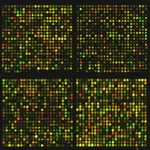
Microarray technology is a high-throughput method used to analyze gene expression levels in a sample. It involves the use of a glass slide or other solid support onto which thousands of known gene sequences are spotted in an array. The sample RNA is labeled with a fluorescent dye and hybridized to the array, allowing for the detection and quantification of gene expression levels.
Microarray technology has several advantages, including its ability to analyze the expression levels of thousands of genes in a single experiment, its high sensitivity, and its ability to detect low-abundance transcripts. However, it also has some limitations, including its inability to detect novel transcripts or splice variants, its dependence on the availability of known gene sequences, and its susceptibility to background noise and cross-hybridization.
There are several types of microarray platforms available, including cDNA microarrays, oligonucleotide microarrays, and bead-based microarrays. Each platform has its own advantages and limitations, and the choice of platform depends on the specific research question and experimental design.
Microarray technology has numerous applications in biomedical research, including the identification of differentially expressed genes between healthy and diseased states, the analysis of gene regulation and methylation, and the study of gene function and regulation. However, it has been largely replaced by RNA sequencing (RNA-Seq) in recent years due to its higher accuracy, sensitivity, and ability to detect novel transcripts and splice variants.
In summary, microarray technology is a high-throughput method used to analyze gene expression levels in a sample. It involves the use of a glass slide or other solid support onto which thousands of known gene sequences are spotted in an array. While it has several advantages, it also has some limitations, and has been largely replaced by RNA sequencing in recent years.
Microarray Technology: Principles and Procedures
Microarray technology is a high-throughput method used to analyze gene expression levels in a sample. It involves the use of a glass slide or other solid support onto which thousands of known gene sequences are spotted in an array. The sample RNA is labeled with a fluorescent dye and hybridized to the array, allowing for the detection and quantification of gene expression levels.
The procedure for microarray analysis involves several steps:
- Sample preparation: The RNA from the sample is extracted and converted to cDNA using reverse transcriptase. The cDNA is then labeled with a fluorescent dye, such as Cy3 or Cy5.
- Hybridization: The labeled cDNA is then hybridized to the microarray, allowing for the detection and quantification of gene expression levels.
- Scanning and data analysis: The microarray is scanned using a laser scanner, and the resulting image is analyzed using specialized software to determine the intensity of the fluorescence for each spot on the array. The intensity of the fluorescence is proportional to the amount of RNA present in the sample.
Microarray Technology: Advantages and Limitations
Advantages of microarray technology include its ability to analyze the expression levels of thousands of genes in a single experiment, its high sensitivity, and its ability to detect low-abundance transcripts. Additionally, microarray technology can provide a snapshot of gene expression levels at a specific point in time, allowing for the analysis of changes in gene expression in response to various stimuli.
However, microarray technology also has some limitations. For example, it is limited to the analysis of known gene sequences, and cannot detect novel transcripts or splice variants. Additionally, microarray technology can be susceptible to background noise and cross-hybridization, which can lead to false positive or false negative results. Finally, microarray technology can be expensive and requires specialized equipment and expertise to perform and analyze the data.
In summary, microarray technology is a high-throughput method used to analyze gene expression levels in a sample. It involves the use of a glass slide or other solid support onto which thousands of known gene sequences are spotted in an array. While microarray technology has several advantages, including its ability to analyze the expression levels of thousands of genes in a single experiment and its high sensitivity, it also has some limitations, such as its inability to detect novel transcripts or splice variants and its susceptibility to background noise and cross-hybridization.
High-Throughput RNA Sequencing (RNA-Seq)
High-throughput RNA sequencing (RNA-Seq) is a powerful technology used for transcriptome analysis. It allows for the sequencing of millions of RNA molecules in a single run, providing a comprehensive view of gene expression levels in a sample. RNA-Seq has several advantages over microarray technology, including its ability to detect novel transcripts and splice variants, its higher accuracy and dynamic range, and its ability to analyze gene regulation and methylation.
The procedure for RNA-Seq involves several steps, including RNA extraction, library preparation, sequencing, and data analysis. RNA is extracted from the sample and converted to cDNA, which is then amplified and prepared into a sequencing library. The library is then sequenced using a high-throughput sequencing platform, such as Illumina or PacBio. The resulting sequencing reads are then mapped to a reference genome or transcriptome, and gene expression levels are quantified using specialized software.
RNA-Seq has numerous applications in biomedical research, including the identification of differentially expressed genes between healthy and diseased states, the analysis of gene regulation and methylation, and the study of gene function and regulation. It has been widely used in cancer research, allowing for the analysis of intratumor expression heterogeneity and the identification of the molecular basis of formation of many oncological diseases.
However, RNA-Seq also has some limitations. For example, it can be expensive and requires specialized equipment and expertise to perform and analyze the data. Additionally, RNA-Seq can be susceptible to biases in library preparation and sequencing, which can affect the accuracy of gene expression quantification.
In summary, High-throughput RNA sequencing (RNA-Seq) is a powerful technology used for transcriptome analysis, providing a comprehensive view of gene expression levels in a sample. While it has several advantages, including its ability to detect novel transcripts and splice variants and its higher accuracy and dynamic range, it also has some limitations, such as its expense and susceptibility to biases in library preparation and sequencing.
High-Throughput RNA Sequencing (RNA-Seq)
- Principles and Procedures:
RNA-Seq is a recently developed approach to transcriptome profiling that uses deep-sequencing technologies. The procedure for RNA-Seq involves several steps, including RNA extraction, library preparation, sequencing, and data analysis. RNA is extracted from the sample and converted to cDNA, which is then amplified and prepared into a sequencing library. The library is then sequenced using a high-throughput sequencing platform, such as Illumina or PacBio. The resulting sequencing reads are then mapped to a reference genome or transcriptome, and gene expression levels are quantified using specialized software.
- Advantages and Limitations:
Advantages of RNA-Seq include its ability to detect novel transcripts and splice variants, its higher accuracy and dynamic range, and its ability to analyze gene regulation and methylation. RNA-Seq provides a more precise measurement of levels of transcripts and their isoforms than other methods. It can also reveal the precise location of transcription boundaries, to a single-base resolution, and can provide information about how two exons are connected. RNA-Seq can also reveal sequence variations, such as SNPs, in the transcribed regions.
However, RNA-Seq also has some limitations. For example, it can be expensive and requires specialized equipment and expertise to perform and analyze the data. Additionally, RNA-Seq can be susceptible to biases in library preparation and sequencing, which can affect the accuracy of gene expression quantification. The large amount of data generated by RNA-Seq can also be challenging to analyze and interpret, requiring specialized bioinformatics tools and expertise. Furthermore, RNA-Seq may not be suitable for samples with low RNA yield, such as from fine-needle aspirates or single cells, due to the requirement for amplification of the cDNA library.
Comparison of RNA-Seq and Microarray Methodologies
RNA-Seq and microarray are two commonly used methods for transcriptome analysis, each with its own advantages and limitations. Here are some key differences between the two methods:
- Sensitivity and Dynamic Range: RNA-Seq has a higher dynamic range and sensitivity compared to microarray. RNA-Seq can detect low-abundance transcripts and quantify gene expression levels over a wider range, while microarray is limited by the intensity of the fluorescent signal and can only detect transcripts above a certain threshold.
- Detection of Novel Transcripts: RNA-Seq can detect novel transcripts and splice variants, while microarray is limited to the analysis of known gene sequences.
- Cost: RNA-Seq can be more expensive than microarray, particularly for large-scale studies, due to the need for specialized equipment and bioinformatics analysis.
- Data Analysis: RNA-Seq generates a large amount of data, requiring specialized bioinformatics tools and expertise for data analysis, while microarray data analysis is more straightforward and can be performed using standard software.
- Reproducibility: RNA-Seq has been shown to have higher reproducibility compared to microarray, particularly for low-abundance transcripts.
- Bias: RNA-Seq can be susceptible to biases in library preparation and sequencing, while microarray can be susceptible to biases in probe design and hybridization.
- Throughput: RNA-Seq can analyze a larger number of samples and replicates compared to microarray, allowing for more robust statistical analysis.
In summary, RNA-Seq and microarray are two commonly used methods for transcriptome analysis, each with its own advantages and limitations. RNA-Seq has a higher dynamic range and sensitivity, can detect novel transcripts and splice variants, and has higher reproducibility compared to microarray. However, RNA-Seq can be more expensive and requires specialized equipment and bioinformatics analysis. Microarray is less expensive and has simpler data analysis, but has a lower dynamic range and sensitivity, and is limited to the analysis of known gene sequences. The choice of method depends on the specific research question and experimental design.
Data Analysis in Transcriptome Experiments
Alignment to Reference
Alignment to a reference genome or transcriptome is a critical step in the analysis of RNA-Seq data. The sequencing reads are aligned to a reference genome or transcriptome to identify the location of the transcripts in the genome, quantify gene expression levels, and detect sequence variations.
The alignment process involves mapping the sequencing reads to the reference genome or transcriptome using specialized software, such as STAR, TopHat, or HISAT2. The alignment software uses various algorithms to identify the best match between the sequencing reads and the reference genome or transcriptome, taking into account factors such as sequence similarity, read quality, and splice site information.
The alignment process can be challenging due to the presence of sequencing errors, genetic variations, and repetitive regions in the genome. To address these challenges, various strategies have been developed, such as using multiple aligners, adjusting alignment parameters, and using reference genomes or transcriptomes with higher accuracy and completeness.
Once the sequencing reads are aligned to the reference genome or transcriptome, the gene expression levels can be quantified using specialized software, such as HTSeq, featureCounts, or StringTie. The quantification process involves counting the number of reads that map to each gene or transcript, taking into account factors such as read length, sequencing depth, and gene length.
In summary, alignment to a reference genome or transcriptome is a critical step in the analysis of RNA-Seq data. The sequencing reads are aligned to the reference genome or transcriptome using specialized software, and the gene expression levels are quantified using specialized software. The alignment process can be challenging due to the presence of sequencing errors, genetic variations, and repetitive regions in the genome. Various strategies have been developed to address these challenges, such as using multiple aligners, adjusting alignment parameters, and using reference genomes or transcriptomes with higher accuracy and completeness.
Quantification of Gene Expression Levels
Quantification of gene expression levels is a critical step in the analysis of RNA-Seq data. The goal of gene expression quantification is to estimate the abundance of each transcript in the sample, which can provide insights into the biological processes and functions of the cells or tissues being studied.
The quantification process involves counting the number of reads that map to each gene or transcript, taking into account factors such as read length, sequencing depth, and gene length. The counts are then normalized to account for differences in sequencing depth and gene length, and the resulting expression levels are used for downstream analysis, such as differential expression analysis, pathway analysis, and functional enrichment analysis.
There are several methods for quantifying gene expression levels from RNA-Seq data, including:
- Count-based methods: These methods involve counting the number of reads that map to each gene or transcript, and normalizing the counts to account for differences in sequencing depth and gene length. Examples of count-based methods include HTSeq, featureCounts, and StringTie.
- Probabilistic methods: These methods involve estimating the probability of observing a given number of reads for each gene or transcript, taking into account factors such as sequencing depth, gene length, and read quality. Examples of probabilistic methods include RSEM, eXpress, and Sailfish.
- Isolation of specific transcripts: These methods involve isolating specific transcripts, such as mRNA or lncRNA, and quantifying their expression levels using targeted sequencing approaches, such as PCR or hybridization capture. Examples of isolation-based methods include qPCR, NanoString, and RNA-CaptureSeq.
The choice of quantification method depends on the specific research question and experimental design. Count-based methods are widely used due to their simplicity and accuracy, while probabilistic methods can provide more accurate estimates of gene expression levels, particularly for low-abundance transcripts. Isolation-based methods can provide more specific information about the expression levels of certain transcripts, but are limited by their lower throughput and higher cost.
In summary, quantification of gene expression levels is a critical step in the analysis of RNA-Seq data. The goal of gene expression quantification is to estimate the abundance of each transcript in the sample, which can provide insights into the biological processes and functions of the cells or tissues being studied. There are several methods for quantifying gene expression levels from RNA-Seq data, including count-based methods, probabilistic methods, and isolation-based methods. The choice of quantification method depends on the specific research question and experimental design.
Novel Transcript and Fusion Gene Identification
To identify novel transcripts and fusion genes in transcriptome analysis, several bioinformatic tools and methods can be used. These methods can be broadly classified into two categories: mapping-first approaches and assembly-first approaches.
Mapping-first approaches involve aligning RNA-seq reads to a reference genome or transcriptome to identify discordantly mapping reads that are suggestive of rearrangements and chimeric read alignments. Examples of such methods include STAR-Fusion, TopHat-Fusion, and MapSplice.
Assembly-first approaches, on the other hand, directly assemble reads into longer transcript sequences followed by identification of chimeric transcripts consistent with chromosomal rearrangements. Examples of such methods include TrinityFusion, SOAPfuse, and JAFFA-Assembly.
The accuracy of these methods can vary, and several studies have been conducted to benchmark their performance. For example, a study published in Genome Biology in 2019 compared 23 different methods for fusion transcript detection, including STAR-Fusion and TrinityFusion, which were developed by the authors. The study found that STAR-Fusion, Arriba, and STAR-SEQR were the most accurate and fastest for fusion detection on cancer transcriptomes.
Another study published in BMC Bioinformatics in 2018 compared six different methods for fusion gene detection, including STAR-Fusion, EricScript, and JAFFA. The study found that STAR-Fusion had the highest precision and recall, followed by EricScript and JAFFA.
It is important to note that the accuracy of these methods can depend on several factors, including the quality and depth of the sequencing data, the complexity of the transcriptome, and the presence of genetic variations or sequencing errors. Therefore, it is recommended to use multiple methods and validate the results using experimental approaches, such as RT-PCR or RNA-seq of additional samples.
In summary, to identify novel transcripts and fusion genes in transcriptome analysis, several bioinformatic tools and methods can be used, including mapping-first and assembly-first approaches. The accuracy of these methods can vary, and it is recommended to use multiple methods and validate the results experimentally.
Data Deposition and Transcriptome Databases
Data deposition and transcriptome databases are important aspects of transcriptome analysis. After performing transcriptome analysis, researchers can deposit their data in public databases, such as Gene Expression Omnibus (GEO) or ArrayExpress, to make their data accessible to other researchers. This allows for data sharing, reproducibility, and reuse of data, which can lead to new discoveries and insights.
Transcriptome databases are also essential tools for transcriptome analysis. These databases contain transcriptome data from various organisms, tissues, and conditions, which can be used for comparative analysis, validation, and hypothesis generation. Examples of transcriptome databases include GEO, ArrayExpress, and the Sequence Read Archive (SRA) at NCBI.
In addition to data deposition and access, transcriptome databases also provide tools for data analysis and visualization. For example, GEO provides tools for data normalization, differential expression analysis, and pathway analysis. These tools can help researchers identify differentially expressed genes, enriched pathways, and functional categories, which can provide insights into the biological processes and functions of the cells or tissues being studied.
In summary, data deposition and transcriptome databases are important aspects of transcriptome analysis. Researchers can deposit their data in public databases to make their data accessible to other researchers, and transcriptome databases provide transcriptome data from various organisms, tissues, and conditions, which can be used for comparative analysis, validation, and hypothesis generation. Transcriptome databases also provide tools for data analysis and visualization, which can help researchers identify differentially expressed genes, enriched pathways, and functional categories.
Data Analysis Software and Algorithms
Data analysis software and algorithms used in transcriptome analysis include tools for quality assessment, alignment, normalization, quantification, and differential expression analysis. Quality assessment tools, such as FastQC and Trim Galore, are used to assess the quality of raw data and filter out low-quality sequences or bases. Alignment tools, such as STAR, TopHat, and HISAT2, align sequenced reads to a reference genome or transcriptome. Normalization tools, such as DESeq2 and edgeR, are used to adjust for differences in sequencing depth and gene length. Quantification tools, such as HTSeq and featureCounts, count the number of reads that map to each gene or transcript. Differential expression tools, such as Cufflinks and Ballgown, identify genes or transcripts that are differentially expressed between samples. De novo transcriptome assembly tools, such as Trinity and Oases, are used to assemble transcriptomes without a reference genome. Fusion gene detection tools, such as STAR-Fusion and EricScript, identify fusion genes in transcriptome data.
Graphical Representations of Transcriptome Analysis Data
Graphical representations of transcriptome analysis data can help researchers visualize and interpret the results of their analysis. Common graphical representations include:
- Heatmaps: Heatmaps are used to visualize the expression levels of genes or transcripts across multiple samples. The expression levels are represented as a color gradient, with red indicating high expression and green indicating low expression. Heatmaps can help researchers identify clusters of genes or transcripts with similar expression patterns.
- Volcano plots: Volcano plots are used to visualize the results of differential expression analysis. The x-axis represents the log2 fold change, and the y-axis represents the negative log10 of the p-value. Genes or transcripts that are significantly differentially expressed are plotted as points, with red indicating up-regulation and blue indicating down-regulation. Volcano plots can help researchers identify genes or transcripts that are significantly differentially expressed between two conditions.
- Principal Component Analysis (PCA) plots: PCA plots are used to visualize the similarity or dissimilarity of samples based on their gene expression profiles. The x-axis and y-axis represent the first and second principal components, respectively, which capture the most variation in the data. Samples that are similar in their gene expression profiles cluster together, while samples that are dissimilar are separated. PCA plots can help researchers identify outliers or batch effects in their data.
- Venn diagrams: Venn diagrams are used to visualize the overlap of differentially expressed genes or transcripts between two or more conditions. The intersections of the circles represent the number of genes or transcripts that are shared between the conditions, while the non-overlapping regions represent the unique genes or transcripts in each condition. Venn diagrams can help researchers identify genes or transcripts that are specific to certain conditions.
- Network diagrams: Network diagrams are used to visualize the interactions between genes or transcripts. The nodes represent the genes or transcripts, and the edges represent the interactions between them. Network diagrams can help researchers identify key genes or transcripts that are central to certain biological processes or pathways.
In summary, graphical representations of transcriptome analysis data can help researchers visualize and interpret the results of their analysis. Common graphical representations include heatmaps, volcano plots, PCA plots, Venn diagrams, and network diagrams. These representations can help researchers identify clusters of genes or transcripts with similar expression patterns, genes or transcripts that are significantly differentially expressed, similarities or dissimilarities between samples, genes or transcripts that are specific to certain conditions, and key genes or transcripts that are central to certain biological processes or pathways.
Applications of Transcriptome Analysis
Transcriptome analysis has the potential to aid in disease diagnosis and biomarker discovery. By analyzing the transcriptome of diseased and healthy samples, researchers can identify genes or transcripts that are differentially expressed between the two conditions. These differentially expressed genes or transcripts can serve as potential biomarkers for the disease.
Biomarkers are measurable indicators of a biological state or condition. In the context of disease, biomarkers can be used for diagnosis, prognosis, or monitoring of disease progression. Transcriptome-based biomarkers can be particularly useful for diseases that lack reliable diagnostic tests or for which existing tests are invasive or expensive.
For example, transcriptome analysis has been used to identify biomarkers for cancer, neurodegenerative diseases, and infectious diseases. In cancer, transcriptome analysis has identified genes or transcripts that are differentially expressed between cancerous and normal tissues, which can be used for early detection, prognosis, and monitoring of cancer progression. In neurodegenerative diseases, transcriptome analysis has identified genes or transcripts that are associated with disease progression, which can be used for early diagnosis and monitoring of disease progression. In infectious diseases, transcriptome analysis has identified genes or transcripts that are associated with host response to infection, which can be used for diagnosis and monitoring of infection.
However, it is important to note that the use of transcriptome-based biomarkers for disease diagnosis and monitoring is still in its infancy, and more research is needed to validate and standardize these biomarkers. Additionally, the use of transcriptome-based biomarkers for disease diagnosis and monitoring is subject to regulatory approval and clinical validation.
In summary, transcriptome analysis has the potential to aid in disease diagnosis and biomarker discovery. By analyzing the transcriptome of diseased and healthy samples, researchers can identify genes or transcripts that are differentially expressed between the two conditions, which can serve as potential biomarkers for the disease. Transcriptome-based biomarkers can be particularly useful for diseases that lack reliable diagnostic tests or for which existing tests are invasive or expensive. However, more research is needed to validate and standardize these biomarkers, and their use for disease diagnosis and monitoring is subject to regulatory approval and clinical validation.
Risk Assessment of New Drugs or Environmental Chemicals
Transcriptome analysis can also be used for risk assessment of new drugs or environmental chemicals. By analyzing the transcriptome of cells or tissues exposed to different concentrations of a chemical, researchers can identify genes or transcripts that are affected by the chemical. These genes or transcripts can provide insights into the mechanism of action of the chemical and its potential toxicity.
For example, transcriptome analysis has been used to assess the toxicity of drugs, pesticides, and industrial chemicals. In drug development, transcriptome analysis can identify genes or transcripts that are affected by a drug, which can help predict its potential side effects and toxicity. In environmental chemistry, transcriptome analysis can identify genes or transcripts that are affected by environmental chemicals, which can help predict their potential health risks and inform regulatory decisions.
Transcriptome analysis can also be used to identify biomarkers of exposure to environmental chemicals. By analyzing the transcriptome of individuals exposed to different levels of a chemical, researchers can identify genes or transcripts that are associated with exposure, which can be used for biomonitoring and risk assessment.
However, it is important to note that the use of transcriptome analysis for risk assessment of new drugs or environmental chemicals is still in its infancy, and more research is needed to validate and standardize the use of transcriptome-based biomarkers. Additionally, the use of transcriptome-based biomarkers for risk assessment is subject to regulatory approval and clinical validation.
In summary, transcriptome analysis can be used for risk assessment of new drugs or environmental chemicals. By analyzing the transcriptome of cells or tissues exposed to different concentrations of a chemical, researchers can identify genes or transcripts that are affected by the chemical, which can provide insights into its mechanism of action and potential toxicity. Transcriptome analysis can also be used to identify biomarkers of exposure to environmental chemicals, which can be used for biomonitoring and risk assessment. However, more research is needed to validate and standardize the use of transcriptome-based biomarkers, and their use for risk assessment is subject to regulatory approval and clinical validation.
Cancer Classification, Pathogenesis Mechanisms, and Outcome Prediction
Transcriptome analysis has been widely used in cancer research to classify tumors, understand the mechanisms of cancer pathogenesis, and predict patient outcomes. By analyzing the gene expression profiles of tumor cells, researchers can identify patterns of gene expression that are specific to certain types of cancer, which can aid in cancer classification and diagnosis. Transcriptome analysis can also reveal the molecular mechanisms underlying cancer pathogenesis, such as the activation of oncogenes and the inactivation of tumor suppressor genes. Additionally, transcriptome analysis can identify genes that are associated with patient outcomes, such as survival or response to treatment, which can inform prognosis and treatment decisions.
Single-cell RNA sequencing (scRNA-seq) is a powerful tool for transcriptome analysis in cancer research. It can reveal the heterogeneity of tumor cells and monitor the progress of tumor development, thereby preventing further cellular deterioration. Furthermore, the transcriptome analysis of immune cells in tumor tissue can be used to classify immune cells, their immune escape mechanisms and drug resistance mechanisms, and to develop effective clinical targeted therapies combined with immunotherapy. Additionally, scRNA-seq enables the study of intercellular communication and the interaction of tumor cells and non-malignant cells to reveal their role in carcinogenesis.
Transcriptome analysis can also be used for risk assessment of new drugs or environmental chemicals in the context of cancer. By analyzing the transcriptome of cells or tissues exposed to different concentrations of a chemical, researchers can identify genes or transcripts that are affected by the chemical, which can provide insights into its mechanism of action and potential toxicity. Transcriptome analysis can also be used to identify biomarkers of exposure to environmental chemicals, which can be used for biomonitoring and risk assessment.
In summary, transcriptome analysis has been widely used in cancer research to classify tumors, understand the mechanisms of cancer pathogenesis, and predict patient outcomes. Single-cell RNA sequencing (scRNA-seq) is a powerful tool for transcriptome analysis in cancer research, revealing the heterogeneity of tumor cells and monitoring the progress of tumor development. Transcriptome analysis can also be used for risk assessment of new drugs or environmental chemicals in the context of cancer.
Personalized Medicine and Individualized Cancer Patient Therapies
Transcriptome analysis has the potential to aid in personalized medicine and individualized cancer patient therapies. By analyzing the gene expression profiles of tumor cells from individual patients, researchers can identify patterns of gene expression that are specific to that patient’s tumor, which can inform personalized treatment decisions. Transcriptome analysis can also reveal the molecular mechanisms underlying the patient’s tumor, such as the activation of oncogenes and the inactivation of tumor suppressor genes, which can inform the selection of targeted therapies.
For example, transcriptome analysis has been used to identify biomarkers of response to certain cancer therapies, such as chemotherapy or targeted therapy. By analyzing the gene expression profiles of tumor cells before and after treatment, researchers can identify genes or transcripts that are associated with response or resistance to treatment. These biomarkers can be used to predict treatment response and inform personalized treatment decisions.
Transcriptome analysis can also be used to monitor the progress of cancer treatment and identify early signs of treatment resistance. By analyzing the gene expression profiles of tumor cells during treatment, researchers can identify changes in gene expression that are associated with treatment resistance, which can inform the selection of alternative therapies.
In summary, transcriptome analysis has the potential to aid in personalized medicine and individualized cancer patient therapies. By analyzing the gene expression profiles of tumor cells from individual patients, researchers can identify patterns of gene expression that are specific to that patient’s tumor, which can inform personalized treatment decisions. Transcriptome analysis can also reveal the molecular mechanisms underlying the patient’s tumor, such as the activation of oncogenes and the inactivation of tumor suppressor genes, which can inform the selection of targeted therapies. Transcriptome analysis can also be used to monitor the progress of cancer treatment and identify early signs of treatment resistance.
Molecular Characterization of Organisms and Tissues at Various Stages of Development
Molecular characterization of organisms and tissues at various stages of development can be achieved through transcriptome analysis. This involves studying the complete set of RNA transcripts produced by the genome under specific circumstances or in a specific cell using high-throughput methods. Transcriptome analysis can help understand the processes of cellular differentiation or embryonic development, and identify targets for treatment.
Transcriptome analysis can be performed using microarray technology or high-throughput RNA sequencing (RNA-Seq). Microarray technology uses a set of defined sequences arranged on a solid substrate, while RNA-Seq detects all transcripts in a sample, including regulatory siRNA and lncRNA transcripts. RNA-Seq requires much less starting material and can measure both low-abundance and high-abundance RNAs over a wide range. It can also identify alternative splicing, novel transcripts, and fusion genes.
Improved sequencing technologies have necessitated improved data analysis methods to deal with the increased volume of data produced by each transcriptome experiment. The results are deposited into transcriptome databases, which are essential tools for transcriptome analysis.
Transcriptome analyses are often presented graphically as heat maps, which represent different levels of expression of given genes in different samples, or Venn diagrams, which count the transcripts which are equivalently regulated in multiple samples.
In summary, molecular characterization of organisms and tissues at various stages of development can be achieved through transcriptome analysis, which involves studying the complete set of RNA transcripts produced by the genome under specific circumstances or in a specific cell using high-throughput methods. Transcriptome analysis can help understand the processes of cellular differentiation or embryonic development, and identify targets for treatment. Improved sequencing technologies and data analysis methods have made transcriptome analysis a powerful tool for molecular characterization.
Identification of Targets for Treatment
Transcriptome analysis can be used to identify targets for treatment in various diseases, including cancer, neurological disorders, and infectious diseases. By analyzing the gene expression profiles of diseased cells or tissues, researchers can identify genes or transcripts that are differentially expressed between healthy and diseased states. These differentially expressed genes or transcripts can be potential targets for treatment.
For example, in cancer research, transcriptome analysis can identify genes or transcripts that are overexpressed or underexpressed in cancer cells compared to normal cells. These genes or transcripts can be potential targets for cancer therapy, such as drugs that target oncogenes or inhibit tumor growth.
In neurological disorders, transcriptome analysis can identify genes or transcripts that are associated with the disease, such as genes involved in neurodegeneration or neuroinflammation. These genes or transcripts can be potential targets for treatment, such as drugs that target neuroinflammation or promote neuroprotection.
In infectious diseases, transcriptome analysis can identify genes or transcripts that are involved in the host response to infection. These genes or transcripts can be potential targets for treatment, such as drugs that modulate the host response or inhibit pathogen replication.
Transcriptome analysis can also be used to identify biomarkers of disease, which can be used for early diagnosis and monitoring of disease progression. Biomarkers can be identified by comparing the gene expression profiles of healthy and diseased samples, and selecting genes or transcripts that are differentially expressed between the two groups.
In summary, transcriptome analysis can be used to identify targets for treatment in various diseases, including cancer, neurological disorders, and infectious diseases. By analyzing the gene expression profiles of diseased cells or tissues, researchers can identify genes or transcripts that are differentially expressed between healthy and diseased states, which can be potential targets for treatment. Transcriptome analysis can also be used to identify biomarkers of disease, which can be used for early diagnosis and monitoring of disease progression.
Case Studies in Transcriptome Analysis
A. Example 1: Serum Stimulation in Cultured Fibroblasts
In this study, researchers used transcriptome analysis to investigate the effects of serum stimulation on cultured fibroblasts. They performed RNA sequencing on fibroblasts that were untreated or treated with serum for different time periods. The analysis revealed that serum stimulation induced significant changes in gene expression, with the majority of differentially expressed genes being upregulated. The most significant changes in gene expression were observed at early time points after serum stimulation. Pathway enrichment analysis revealed that serum stimulation activated several signaling pathways, including the MAPK signaling pathway, the PI3K-Akt signaling pathway, and the Rap1 signaling pathway. The study provides insights into the molecular mechanisms underlying the response of fibroblasts to serum stimulation and highlights the potential of transcriptome analysis in understanding cellular responses to external stimuli.
B. Example 2: Cancer Transcriptome Analysis
Transcriptome analysis has been widely used in cancer research to identify differentially expressed genes and pathways between cancerous and normal tissues. In this study, researchers performed RNA sequencing on breast cancer tissues and normal breast tissues. The analysis revealed that breast cancer tissues had significantly altered gene expression profiles compared to normal breast tissues. Several genes and pathways were found to be differentially expressed, including genes involved in cell cycle regulation, DNA damage response, and immune response. The study provides insights into the molecular mechanisms underlying breast cancer and highlights the potential of transcriptome analysis in identifying biomarkers and therapeutic targets for cancer.
C. Example 3: Transcriptome Analysis in Skin and Dermatology
Transcriptome analysis has also been used in skin and dermatology research to investigate the molecular mechanisms underlying skin disorders and identify potential therapeutic targets. In this study, researchers performed RNA sequencing on skin samples from patients with psoriasis and healthy controls. The analysis revealed that psoriasis skin had significantly altered gene expression profiles compared to healthy skin. Several genes and pathways were found to be differentially expressed, including genes involved in inflammation, immune response, and epidermal differentiation. The study provides insights into the molecular mechanisms underlying psoriasis and highlights the potential of transcriptome analysis in identifying biomarkers and therapeutic targets for skin disorders.
In summary, transcriptome analysis has been widely used in various fields of biomedical research, including serum stimulation in cultured fibroblasts, cancer, and skin and dermatology. The analysis can reveal differentially expressed genes and pathways between different conditions, providing insights into the molecular mechanisms underlying various biological processes and diseases. The results can also identify potential biomarkers and therapeutic targets, highlighting the potential of transcriptome analysis in personalized medicine and individualized patient therapies.
Future Perspectives and Challenges in Transcriptome Analysis
The future perspectives and challenges in transcriptome analysis include emerging technologies and methodologies, ethical considerations and regulations, and interdisciplinary collaboration and data sharing.
A. Emerging Technologies and Methodologies:
- Single-cell sequencing: Single-cell sequencing technology has revolutionized transcriptome analysis by enabling the measurement of gene expression at the single-cell level. This technology has the potential to reveal cell-to-cell heterogeneity and provide insights into the dynamics of tissue and organism development. However, it also poses unique data science problems, such as the analysis of vast quantities of data and the need for scalable data analysis models and methods.
- Spatial transcriptomics: Spatial transcriptomics is an emerging technology that allows for the measurement of gene expression in specific locations within tissues. This technology has the potential to reveal the spatial organization of cells and tissues and provide insights into the interactions between cells. However, it also poses challenges in data analysis and interpretation.
- Long-read sequencing: Long-read sequencing technology has the potential to reveal full-length transcripts and provide insights into alternative splicing and gene fusion events. However, it also poses challenges in data analysis and interpretation due to the higher error rate and the need for specialized bioinformatics tools.
B. Ethical Considerations and Regulations:
- Privacy concerns: Transcriptome analysis can reveal sensitive information about individuals, such as their health status and genetic makeup. Therefore, there are ethical considerations and regulations regarding the use and sharing of transcriptome data.
- Data security: Transcriptome data can be sensitive and personal, and therefore, there are regulations regarding data security and privacy.
- Informed consent: Transcriptome analysis often involves the use of human samples, and therefore, there are regulations regarding informed consent and ethical considerations regarding the use of human samples.
C. Interdisciplinary Collaboration and Data Sharing:
- Collaboration between biologists and data scientists: Transcriptome analysis involves the integration of biology and data science, and therefore, there is a need for collaboration between biologists and data scientists.
- Data sharing: Transcriptome data can be large and complex, and therefore, there is a need for data sharing and standardization to enable collaboration and reproducibility.
- Open science: Transcriptome analysis can benefit from open science practices, such as open data and open source software, to enable collaboration and reproducibility.
In summary, the future perspectives and challenges in transcriptome analysis include emerging technologies and methodologies, ethical considerations and regulations, and interdisciplinary collaboration and data sharing. Emerging technologies such as single-cell sequencing, spatial transcriptomics, and long-read sequencing have the potential to reveal new insights into biological systems but also pose unique data analysis challenges. Ethical considerations and regulations regarding privacy, data security, and informed consent are important to ensure the responsible use of transcriptome data. Interdisciplinary collaboration and data sharing are essential to enable collaboration, reproducibility, and the advancement of transcriptome analysis.
Conclusion
In conclusion, transcriptome analysis is a powerful tool in biomedical research that has the potential to reveal insights into the molecular mechanisms underlying various biological processes and diseases. It can identify differentially expressed genes and pathways between different conditions, providing a better understanding of the underlying biology. Transcriptome analysis can also identify potential biomarkers and therapeutic targets, highlighting the potential of transcriptome analysis in personalized medicine and individualized patient therapies.
Emerging technologies such as single-cell sequencing, spatial transcriptomics, and long-read sequencing have the potential to reveal new insights into biological systems but also pose unique data analysis challenges. Ethical considerations and regulations regarding privacy, data security, and informed consent are important to ensure the responsible use of transcriptome data. Interdisciplinary collaboration and data sharing are essential to enable collaboration, reproducibility, and the advancement of transcriptome analysis.
Transcriptome analysis has the potential to revolutionize our understanding of biology and disease, and further study and exploration in the field are encouraged. The development of new technologies and methodologies, the integration of transcriptome analysis with other -omics approaches, and the application of transcriptome analysis in personalized medicine and individualized patient therapies are exciting areas of research that have the potential to advance our understanding of biology and disease. The integration of transcriptome analysis with other -omics approaches, such as genomics, proteomics, and metabolomics, can provide a more comprehensive understanding of biological systems. The application of transcriptome analysis in personalized medicine and individualized patient therapies can lead to the development of more effective and targeted treatments for various diseases.
In summary, transcriptome analysis is a powerful tool in biomedical research that has the potential to reveal insights into the molecular mechanisms underlying various biological processes and diseases. Emerging technologies and methodologies, ethical considerations and regulations, and interdisciplinary collaboration and data sharing are important factors to consider in the advancement of transcriptome analysis. Further study and exploration in the field are encouraged to advance our understanding of biology and disease.
Related posts:
![R-program-data-plot]()
Introductory R and Data Visualization for Bioinformatics - A complete Guide
bioinformatics![bioinformatics-FAQ]()
100 FAQ's Guide to Pursuing a Career in Bioinformatics
bioinformatics![microarray analysis]()
Step-by-Step Guide: Analyzing Microarray Data in Bioconductor
bioinformatics![Futureofbioinformatics]()
The Future of Bioinformatics: Global Perspectives and Challenges
bioinformatics![Bioinformatics research]()
Foundations and Applications of Bioinformatics
bioinformatics![AI-bioinformatics]()
Step-by-Step Guide: Determining a Good Threshold for Log2 Fold Change in Differential Expression Ana...
bioinformatics![Large Language Models]()
A Practical Introduction to Large Language Models in Biomedical Data Science Research
A.I![Bioinformatics for personalized medicine: the synergistic cycle of hypothesis-driven and data-driven experimentation]()
Incorporating Bioinformatics into Undergraduate Education: Practical Approaches and Benefits
bioinformatics![bioinformatics lab]()
RNA-seq Data Analysis: Ultimate Guide from Sample preparation to RAM Requirements
Guides![CRISPR-COVID-19]()
Can Crispr and Precision Medicine Lead to Curing All Diseases?
genomics![linux-basics-commands-fundamentals]()
10 Essential Tips to Master Linux Quickly for Bioinformatics Success
bioinformatics![quantum computer and AI]()
The Best Ways to Learn AI in 2025
A.I![Omics data analysis]()
Guide to Analyzing Protein and Nucleotide FASTA Sequences Using R: From Basics to Advanced Technique...
bioinformatics![Next-Generation Sequencing (NGS) Instruments of 2023]()
Top Next-Generation Sequencing (NGS) Instruments of 2023: A Comparative Guide
bioinformatics![Single-cell Atlases]()
Introduction to Single-cell Atlases
transcriptomics![Aicoursesinindia]()
Artificial Intelligence Courses in India
A.I