microbioloyg-bioinformatics

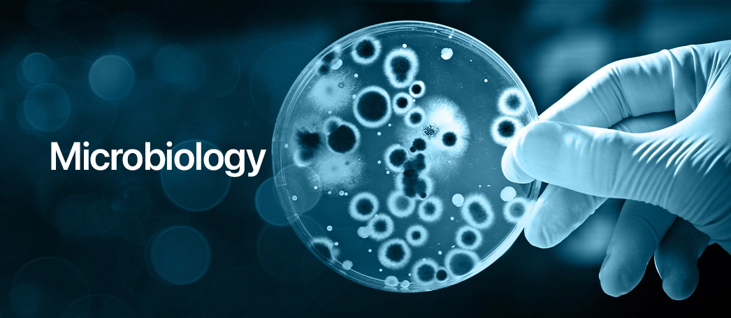
Microbiology: Foundations and Applications in Bioinformatics

March 31, 2024 Off By admin
Shares

Introduction to Microbiology

Scope of Microbiology

Microbiology is a vast field that encompasses the study of microscopic organisms, including bacteria, viruses, fungi, protozoa, and algae. It has numerous branches and applications, including:

  1. Medical Microbiology: Study of microorganisms that cause infectious diseases in humans, including bacteria, viruses, fungi, and parasites. It involves the diagnosis, treatment, and prevention of infectious diseases.
  2. Environmental Microbiology: Study of the roles of microorganisms in the environment, including their roles in nutrient cycling, biodegradation, and environmental impact.
  3. Industrial Microbiology: Application of microorganisms in industrial processes, such as fermentation, waste treatment, bioremediation, and the production of food, beverages, antibiotics, and enzymes.
  4. Agricultural Microbiology: Study of microorganisms in soil fertility, plant nutrition, and plant diseases, with applications in agriculture, horticulture, and forestry.
  5. Food Microbiology: Study of microorganisms in food production, processing, preservation, and safety, including the prevention of foodborne illnesses.
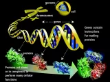
  6. Microbial Genetics: Study of the genetics of microorganisms, including gene expression, genetic transfer, and genetic engineering for biotechnological applications.
  7. Immunology: Study of the immune system, including how it responds to infectious agents and how it can be manipulated for the treatment of diseases.
  8. Microbial Ecology: Study of the interactions between microorganisms and their environments, including their roles in ecosystems, biogeochemical cycles, and climate change.
  9. Veterinary Microbiology: Study of microorganisms that affect animal health, including those that cause diseases in livestock and pets.
  10. Water Microbiology: Study of microorganisms in water, including their roles in water quality, purification, and wastewater treatment.

Microbiology plays a crucial role in various fields, including medicine, agriculture, food production, biotechnology, environmental science, and veterinary science. Its scope continues to expand with advancements in technology and our understanding of microbial life.

History of Microbiology

The history of microbiology dates back to ancient times when people first observed microorganisms without understanding their nature. However, modern microbiology began to emerge in the 17th century with the advent of the microscope and the work of pioneering scientists. Here are some key milestones in the history of microbiology:

  1. Antonie van Leeuwenhoek (1632-1723): A Dutch scientist who is often considered the father of microbiology. He was the first to observe and describe microorganisms, which he called “animalcules,” using a simple microscope of his own design.
  2. Francesco Redi (1626-1697): An Italian physician who conducted experiments to refute the theory of spontaneous generation, which posited that living organisms could arise spontaneously from non-living matter. His work laid the foundation for the field of microbiology.
  3. Louis Pasteur (1822-1895): A French chemist and microbiologist who made several groundbreaking discoveries in the field of microbiology. He is known for his experiments that disproved the theory of spontaneous generation, his development of the process of pasteurization to prevent food spoilage, and his development of vaccines for rabies and anthrax.
  4. Robert Koch (1843-1910): A German physician and microbiologist who is considered one of the founders of modern bacteriology. He developed a series of postulates, known as Koch’s postulates, to establish the causal relationship between a microorganism and a disease. He also identified the causative agents of several diseases, including anthrax, tuberculosis, and cholera.
  5. Joseph Lister (1827-1912): A British surgeon who is known for pioneering antiseptic surgery. He introduced the use of carbolic acid (phenol) as an antiseptic to sterilize surgical instruments and clean wounds, reducing the risk of infection.
  6. Martinus Beijerinck (1851-1931): A Dutch microbiologist who discovered the first virus, tobacco mosaic virus, and coined the term “virus” to describe it. He also made significant contributions to the study of nitrogen fixation and soil microbiology.
  7. Sergei Winogradsky (1856-1953): A Russian microbiologist who is known for his work in environmental microbiology and microbial ecology. He discovered several new species of bacteria and pioneered the study of chemolithotrophy, the process by which microorganisms obtain energy from inorganic compounds.

These pioneering scientists laid the foundation for the field of microbiology, which has since grown to encompass a wide range of disciplines and applications. Today, microbiology plays a crucial role in medicine, agriculture, food production, biotechnology, environmental science, and many other fields.

Spontaneous generation

Spontaneous generation was a widely held belief that living organisms could arise spontaneously from non-living matter. This idea was prevalent in ancient times and was supported by observations such as the appearance of maggots on decaying meat or the sudden appearance of fish in dried-up ponds after rain.

The theory of spontaneous generation was challenged and eventually disproved through a series of experiments by scientists such as Francesco Redi and Louis Pasteur. Redi conducted experiments in the 17th century showing that maggots only appeared on meat when flies were allowed to land on it, refuting the idea that they arose spontaneously from the meat.

Pasteur’s experiments in the 19th century provided further evidence against spontaneous generation. He demonstrated that microorganisms did not spontaneously generate in nutrient broths when exposed to air, but rather were carried in the air and could be prevented from entering the broth by proper sterilization techniques. This led to the development of aseptic techniques and the understanding that microorganisms come from pre-existing microorganisms.

The disproval of spontaneous generation was a significant milestone in the history of microbiology, as it laid the foundation for the modern understanding of the microbial world and the development of microbiological techniques such as sterilization, pasteurization, and aseptic technique.

Germ theory of diseases

The germ theory of disease is the concept that microorganisms, primarily bacteria and viruses, are the cause of many diseases. This theory revolutionized medicine and our understanding of infectious diseases. Here’s an overview of its development and impact:

  1. Antonie van Leeuwenhoek (1632-1723): While Leeuwenhoek’s work predates the formal germ theory, his observations of microorganisms laid the groundwork for understanding their role in disease.
  2. Louis Pasteur (1822-1895): Pasteur’s experiments in the mid-19th century provided strong evidence for the germ theory. He demonstrated that microorganisms are present in the air and can contaminate sterile solutions, leading to spoilage. He also showed that heating liquids to specific temperatures could kill these microorganisms, a process known as pasteurization.
  3. Robert Koch (1843-1910): Koch’s work further solidified the germ theory and provided a framework for identifying the specific microorganisms responsible for particular diseases. He developed a series of postulates (Koch’s postulates) that are used to establish a causal relationship between a microorganism and a disease.
  4. Impact on Medicine: The acceptance of the germ theory revolutionized medicine by providing a scientific basis for understanding and treating infectious diseases. It led to improvements in sanitation, hygiene, and medical practices, such as sterilization and aseptic techniques.
  5. Vaccination: The germ theory also contributed to the development of vaccines. By understanding that microorganisms cause diseases, scientists were able to develop vaccines to prevent infections by stimulating the immune system to recognize and attack specific pathogens.
  6. Antibiotics: The germ theory paved the way for the development of antibiotics, which are substances that can kill or inhibit the growth of bacteria. Antibiotics have been instrumental in treating bacterial infections and saving countless lives.

Overall, the germ theory of disease has had a profound impact on public health and medicine, leading to significant advancements in our ability to prevent, diagnose, and treat infectious diseases.

Cell theory

Cell theory is a fundamental theory in biology that states:

  1. All living organisms are composed of one or more cells.
  2. The cell is the basic unit of structure and organization in organisms.
  3. Cells arise from pre-existing cells through cell division.

The cell theory was proposed in the 19th century and is considered one of the foundations of modern biology. Here’s a brief overview of its development:

  1. Robert Hooke (1635-1703): In 1665, Robert Hooke, an English scientist, discovered cells while examining a thin slice of cork under a microscope. He coined the term “cell” to describe the tiny compartments he observed, which reminded him of the cells inhabited by monks in a monastery.
  2. Antonie van Leeuwenhoek (1632-1723): A Dutch scientist, Leeuwenhoek made significant improvements to the microscope and was the first to observe and describe single-celled organisms, which he called “animalcules.”
  3. Matthias Schleiden (1804-1881) and Theodor Schwann (1810-1882): In the 1830s, Schleiden, a German botanist, and Schwann, a German zoologist, independently formulated the cell theory based on their observations and research. Schleiden concluded that all plant tissues are composed of cells, while Schwann proposed that animals are also made up of cells.
  4. Rudolf Virchow (1821-1902): A German physician, Virchow, later added to the cell theory by stating that cells can only arise from pre-existing cells, challenging the idea of spontaneous generation.

The cell theory laid the groundwork for the field of cell biology, which studies the structure, function, and behavior of cells. It has since been expanded and refined with advancements in technology, leading to a deeper understanding of cellular processes and the development of modern biotechnology and medicine.

Contributions

Antonie van Leeuwenhoek (1632-1723) was a Dutch scientist and tradesman who made pioneering contributions to the field of microscopy and microbiology. His discoveries laid the foundation for the field of microbiology. Some of his key contributions include:

  1. Microscopic Observations: Leeuwenhoek was the first to observe and describe microorganisms, which he called “animalcules.” Using microscopes of his own design, he observed bacteria, protozoa, and other microorganisms for the first time, opening up a new world of the unseen.
  2. Advancements in Microscopy: Leeuwenhoek made significant improvements to the design and construction of microscopes, achieving magnifications of up to 270 times, far surpassing the capabilities of other microscopes of his time.
  3. Discovery of Sperm Cells: Leeuwenhoek was the first to observe and describe sperm cells, which he called “animalcules of the semen,” in human semen and other animals.
  4. Observation of Blood Cells: He was also the first to observe and describe red blood cells, which he called “red globules,” in various animals.
  5. Discovery of Microscopic Structures: Leeuwenhoek’s observations extended beyond microorganisms. He also described and illustrated microscopic structures in a variety of materials, including plants, insects, and minerals.
  6. Scientific Communication: Despite his lack of formal scientific training, Leeuwenhoek communicated his findings to the scientific community through letters to the Royal Society of London, where they were published and widely disseminated, contributing to the advancement of scientific knowledge.

Antonie van Leeuwenhoek’s contributions to microscopy and microbiology were groundbreaking and laid the foundation for the development of these fields. His meticulous observations and innovative approach to microscopy opened up new avenues of scientific inquiry and revolutionized our understanding of the microscopic world.

Here is a brief overview of the contributions of each of these scientists:

  1. Joseph Lister (1827-1912): Known as the father of antiseptic surgery, Lister pioneered the use of antiseptic agents to prevent surgical infections. His work significantly reduced post-operative infections and mortality rates.
  2. Robert Koch (1843-1910): A German physician and microbiologist, Koch is known for his work on identifying the causative agents of anthrax, tuberculosis, and cholera. He developed Koch’s postulates, a series of criteria to establish the causal relationship between a microorganism and a disease.
  3. Louis Pasteur (1822-1895): A French chemist and microbiologist, Pasteur made significant contributions to the fields of microbiology and immunology. He developed the process of pasteurization to prevent food spoilage and the spread of disease. He also developed vaccines for rabies and anthrax.
  4. Edward Jenner (1749-1823): An English physician, Jenner is known for developing the smallpox vaccine. His work laid the foundation for the concept of vaccination and the eventual eradication of smallpox.
  5. John Tyndall (1820-1893): An Irish physicist, Tyndall made important contributions to our understanding of the properties of air and the transmission of light through different substances. He also conducted experiments that demonstrated the presence of heat-resistant microbial spores in the air.
  6. Sergei N. Winogradsky (1856-1953): A Russian microbiologist, Winogradsky is known for his work in environmental microbiology and microbial ecology. He discovered several new species of bacteria and pioneered the study of chemolithotrophy.
  7. Alexander Fleming (1881-1955): A Scottish biologist and pharmacologist, Fleming discovered the antibiotic properties of penicillin. His discovery revolutionized the treatment of bacterial infections and led to the development of many other antibiotics.

Each of these scientists made significant contributions to the fields of microbiology, immunology, and medicine, advancing our understanding of infectious diseases and improving public health.

Microbial cell structure and function

Cell Size, Arrangement, and Shape:

  • Bacterial cells are typically much smaller than eukaryotic cells, ranging from about 0.2 to 2.0 micrometers in diameter.
  • Bacteria can be found in various arrangements, including cocci (spherical), bacilli (rod-shaped), and spirilla (spiral-shaped).
  • The shape of bacterial cells can vary greatly and is often influenced by the cell wall and cytoskeleton.

Capsule, Slime, and Pili:

  • Capsules are protective layers of polysaccharides or proteins that surround some bacterial cells, providing protection against host immune responses.
  • Slime layers are similar to capsules but are more loosely organized and are involved in attachment to surfaces.
  • Pili (or fimbriae) are hair-like appendages that extend from the surface of some bacterial cells and are involved in attachment to surfaces or other cells.

Spores:

  • Spores are specialized, dormant structures formed by some bacteria in response to unfavorable environmental conditions.
  • Spores are highly resistant to heat, desiccation, and chemical agents, allowing the bacteria to survive harsh conditions.
  • Examples of spore-forming bacteria include Bacillus and Clostridium species.

Gram-Negative & Gram-Positive Cell Wall and Membrane:

  • Gram-negative bacteria have a thin peptidoglycan layer in their cell wall and an outer membrane composed of lipopolysaccharides (LPS).
  • Gram-positive bacteria have a thick peptidoglycan layer in their cell wall but lack an outer membrane.
  • The Gram stain is a common laboratory technique used to differentiate between Gram-negative and Gram-positive bacteria based on differences in cell wall structure.

Periplasmic Space:

  • The periplasmic space is a region between the inner and outer membranes of Gram-negative bacteria.
  • It contains a variety of proteins and enzymes involved in nutrient uptake, cell wall synthesis, and detoxification.

Viruses:

  • Viruses are infectious agents that are much smaller than bacteria and consist of genetic material (DNA or RNA) surrounded by a protein coat (capsid).
  • Viruses are obligate intracellular parasites, meaning they can only replicate inside host cells.
  • They infect a wide range of organisms, including animals, plants, fungi, and bacteria.

Mycoplasma:

  • Mycoplasma are bacteria that lack a cell wall, making them resistant to antibiotics that target cell wall synthesis.
  • They are among the smallest known free-living organisms and have a simple cell structure.
  • Mycoplasma species can cause diseases such as pneumonia and urinary tract infections in humans and animals.

Eukaryotic Microbes:

  • Eukaryotic microbes include protists, fungi, and algae, which are single-celled organisms with a complex internal structure.
  • Protists are a diverse group of eukaryotes that include both photosynthetic and non-photosynthetic organisms.
  • Fungi are a group of eukaryotes that include yeasts, molds, and mushrooms. They play important roles in decomposition, nutrient cycling, and food production.
  • Algae are photosynthetic eukaryotes that are important primary producers in aquatic ecosystems and also play a role in carbon fixation and oxygen production.

Microbial Taxonomy

Taxonomy: principle and its types

Taxonomy is the science of classifying and naming organisms based on their similarities and evolutionary relationships. The principle of taxonomy is to organize the diversity of life into hierarchical categories that reflect evolutionary history. The primary goal of taxonomy is to provide a system of classification that is logical, easily understood, and universally applicable.

There are several types of taxonomy, including:

  1. Hierarchical Taxonomy: This type of taxonomy organizes organisms into a hierarchical structure based on their characteristics. The highest level is the domain, followed by kingdom, phylum, class, order, family, genus, and species.
  2. Phenetic Taxonomy: Phenetic taxonomy classifies organisms based on their overall similarity in physical and biochemical characteristics. This approach does not consider evolutionary relationships and relies on observable traits.
  3. Cladistic Taxonomy: Cladistic taxonomy, also known as phylogenetic taxonomy, classifies organisms based on their evolutionary relationships. It uses cladograms, or branching diagrams, to show the relationship between different groups of organisms based on shared derived characteristics.
  4. Numerical Taxonomy: Numerical taxonomy uses statistical methods to analyze large sets of data on characteristics of organisms to classify them. This approach is based on quantitative data and does not rely on subjective assessments of similarity.
  5. Alpha Taxonomy: Alpha taxonomy is concerned with the identification and classification of individual species. It involves describing new species and assigning them to existing taxonomic groups based on their similarities and differences.
  6. Beta Taxonomy: Beta taxonomy deals with the broader classification of organisms beyond the species level. It focuses on the relationships between different species and higher taxonomic groups.
  7. Polyphasic Taxonomy: Polyphasic taxonomy integrates multiple sources of data, including morphology, genetics, ecology, and biochemistry, to classify organisms. This approach provides a more comprehensive understanding of evolutionary relationships.

Taxonomy plays a crucial role in biology by providing a framework for understanding the diversity of life and its evolutionary history. It helps scientists organize and communicate information about organisms, study their relationships, and make predictions about their behavior and characteristics.

Classical approach

The classical approach to taxonomy includes several methods for classifying organisms based on different types of data. These methods include numerical, chemical, serological, and genetic approaches:

  1. Numerical Taxonomy: Numerical taxonomy uses statistical methods to analyze quantitative data on the characteristics of organisms. This approach groups organisms based on similarities in their measurable traits, such as morphology, physiology, or behavior. Cluster analysis and similarity coefficients are commonly used in numerical taxonomy to determine the degree of similarity between organisms and group them accordingly.
  2. Chemical Taxonomy: Chemical taxonomy involves analyzing the chemical composition of organisms to classify them. This can include studying the composition of cell walls, membranes, proteins, lipids, and other biochemical markers. Chemical taxonomy is particularly useful for identifying microorganisms and distinguishing between closely related species.
  3. Serological Taxonomy: Serological taxonomy uses serological techniques, such as immunodiffusion and enzyme-linked immunosorbent assay (ELISA), to study the antigenic properties of organisms. By analyzing the immune response of organisms to specific antigens, serological taxonomy can help classify organisms based on their antigenic similarities and differences.
  4. Genetic Taxonomy: Genetic taxonomy involves studying the genetic material of organisms, such as DNA or RNA, to classify them. This can include analyzing genetic sequences, such as those of specific genes or entire genomes, to determine evolutionary relationships between organisms. Genetic taxonomy is particularly useful for studying the evolutionary history of organisms and identifying genetic markers for classification.

These classical approaches to taxonomy have been widely used in the past and have provided valuable insights into the classification and identification of organisms. However, modern taxonomy increasingly relies on molecular techniques, such as DNA sequencing, for more precise and comprehensive classifications.

Bacterial taxonomy

Bergey’s Manual of Systematic Bacteriology is a renowned reference work in bacterial taxonomy. It provides comprehensive information on the classification, identification, and characterization of bacteria. The manual is organized into several volumes, with each volume focusing on different groups of bacteria.

  1. Volume 1: The Archaea and the Deeply Branching and Phototrophic Bacteria: This volume covers the Archaea domain and includes information on the deeply branching and phototrophic bacteria. It provides detailed descriptions of the taxonomy, morphology, physiology, and ecology of these groups.
  2. Volume 2: The Proteobacteria: This volume focuses on the Proteobacteria, which is a large and diverse group of bacteria that includes many medically, industrially, and ecologically important species. It provides detailed information on the classification, identification, and characterization of Proteobacteria.
  3. Volume 3: The Firmicutes: This volume covers the Firmicutes, a phylum of bacteria that includes many important human pathogens, such as Clostridium difficile and Staphylococcus aureus. It provides comprehensive information on the taxonomy, morphology, physiology, and ecology of Firmicutes.
  4. Volume 4: The Bacteroidetes, Spirochaetes, Tenericutes (Mollicutes), Acidobacteria, Fibrobacteres, Fusobacteria, Dictyoglomi, Gemmatimonadetes, Lentisphaerae, Verrucomicrobia, Chlamydiae, and Planctomycetes: This volume covers a diverse range of bacterial phyla, including the Bacteroidetes, Spirochaetes, Tenericutes, Acidobacteria, Fibrobacteres, Fusobacteria, Dictyoglomi, Gemmatimonadetes, Lentisphaerae, Verrucomicrobia, Chlamydiae, and Planctomycetes. It provides detailed descriptions of the taxonomy, morphology, physiology, and ecology of these groups.

Bergey’s Manual of Systematic Bacteriology is widely used by microbiologists, taxonomists, and researchers as a reference for the classification and identification of bacteria. It is considered a definitive resource in the field of bacterial taxonomy and is regularly updated to reflect advances in the field.

Methods and Techniques in Microbiology

Microscopy

Microscopy is the science of using microscopes to observe objects and details that are too small to be seen with the naked eye. There are several types of microscopes, each with its own principles and applications:

  1. Light Microscope: Also known as an optical microscope, this type of microscope uses visible light to illuminate and magnify samples. It is commonly used in biology, medicine, and materials science.
  2. Phase Contrast Microscope: This type of microscope enhances the contrast of transparent and colorless objects by exploiting differences in the phase of light passing through the sample.
  3. Dark Field Microscope: Dark field microscopy illuminates the sample with light that is scattered by the sample, rather than directly transmitted through it. This creates a bright image of the object against a dark background, making it useful for observing unstained samples.
  4. Bright Field Microscope: This is the most common type of microscope, where the specimen is illuminated with white light, and the image is viewed against a bright background. It is suitable for observing stained or naturally pigmented samples.
  5. Fluorescence Microscope: This type of microscope uses fluorescence to visualize structures within a specimen. Fluorescent molecules in the sample emit light of a different color when illuminated with specific wavelengths, allowing for the visualization of specific structures or molecules.
  6. Interference Microscope (Stereo Microscope): This type of microscope uses interference patterns to enhance the contrast and three-dimensional appearance of the sample. It is commonly used for dissecting and examining larger specimens at low magnifications.
  7. Confocal Microscope: Confocal microscopy uses a pinhole to eliminate out-of-focus light, allowing for the acquisition of sharp, high-contrast images of thick specimens. It is often used in biology and materials science for imaging thick specimens or for 3D imaging.
  8. Electron Microscope (TEM and SEM): Electron microscopes use a beam of electrons to illuminate the specimen, providing much higher magnification and resolution than light microscopes. Transmission Electron Microscopy (TEM) transmits electrons through a thin section of the specimen to create an image, while Scanning Electron Microscopy (SEM) scans a focused electron beam across the surface of the specimen to create a detailed 3D image.

Each type of microscope has its advantages and limitations, and the choice of microscope depends on the specific requirements of the sample and the desired level of detail and resolution.

Nutrition, Growth, and Culturing

Microbiological media

Microbiological media are nutrient-rich substances used to grow and cultivate microorganisms in the laboratory. These media provide the necessary nutrients, pH, and other conditions to support the growth of specific types of microorganisms. There are several types of microbiological media, classified based on their composition and purpose:

  1. Nutrient Agar: Nutrient agar is a general-purpose medium used to grow a wide variety of bacteria and fungi. It contains beef extract, peptone, agar, and water.
  2. Blood Agar: Blood agar is a differential medium used to differentiate bacteria based on their ability to hemolyze (break down) red blood cells. It contains nutrient agar supplemented with sheep or horse blood.
  3. MacConkey Agar: MacConkey agar is a selective and differential medium used to isolate and differentiate Gram-negative bacteria, particularly enteric bacteria. It contains bile salts and crystal violet to inhibit the growth of Gram-positive bacteria, and lactose and pH indicators to differentiate lactose fermenters from non-fermenters.
  4. Sabouraud Agar: Sabouraud agar is used to isolate fungi and yeasts from clinical specimens. It contains a low pH and high glucose concentration to inhibit bacterial growth and promote fungal growth.
  5. Eosin Methylene Blue (EMB) Agar: EMB agar is a selective and differential medium used to isolate and differentiate Gram-negative bacteria, particularly fecal coliforms. It contains eosin and methylene blue dyes to inhibit the growth of Gram-positive bacteria and differentiate lactose fermenters from non-fermenters.
  6. Mannitol Salt Agar: Mannitol salt agar is a selective and differential medium used to isolate and differentiate staphylococci, particularly Staphylococcus aureus, from other bacteria. It contains high salt concentration to select for staphylococci and mannitol and pH indicators to differentiate mannitol fermenters from non-fermenters.
  7. Thioglycollate Broth: Thioglycollate broth is a liquid medium used to culture anaerobic and microaerophilic bacteria. It contains thioglycollate to reduce oxygen tension and resazurin as an oxygen indicator.

These are just a few examples of the many types of microbiological media used in laboratory settings. The choice of medium depends on the specific requirements of the microorganisms being cultured and the goals of the experiment.

Selective and differential media

Selective and differential media are types of microbiological media used to isolate and differentiate specific types of microorganisms based on their growth characteristics. Here’s how they differ:

  1. Selective Media: Selective media contain ingredients that inhibit the growth of certain types of microorganisms while allowing the growth of others. This selectivity is achieved through the inclusion of substances such as antibiotics, dyes, or salts that inhibit the growth of specific groups of bacteria. Selective media are used to isolate and culture specific types of bacteria from complex samples.

    Examples of selective media include:

    • MacConkey agar, which selects for Gram-negative bacteria by inhibiting the growth of Gram-positive bacteria with crystal violet and bile salts.
    • Mannitol salt agar, which selects for staphylococci by incorporating a high concentration of salt that inhibits the growth of other bacteria.
    • Eosin Methylene Blue (EMB) agar, which selects for Gram-negative bacteria by inhibiting the growth of Gram-positive bacteria with eosin and methylene blue dyes.
  2. Differential Media: Differential media contain substances that allow for the differentiation of microorganisms based on their metabolic characteristics. These substances can include pH indicators, sugars, and other compounds that change color or produce visible changes when metabolized by specific bacteria. Differential media are used to identify and characterize bacteria based on their ability to ferment sugars, produce enzymes, or metabolize specific compounds.

    Examples of differential media include:

    • Blood agar, which differentiates bacteria based on their ability to hemolyze (break down) red blood cells. Different patterns of hemolysis can indicate different species or strains of bacteria.
    • MacConkey agar, which not only selects for Gram-negative bacteria but also differentiates lactose-fermenting (pink colonies) from non-fermenting (colorless colonies) bacteria.
    • Triple sugar iron (TSI) agar, which contains sugars and pH indicators to differentiate bacteria based on their ability to ferment sugars and produce gas.

Selective and differential media are valuable tools in microbiology for isolating and identifying specific types of bacteria from complex samples. They are often used in combination to achieve both selectivity and differentiation in bacterial cultures.

Growth curve, growth kinetics

The growth curve and growth kinetics describe the growth pattern of microorganisms in a culture over time. The growth curve typically consists of four phases: lag phase, exponential (log) phase, stationary phase, and death phase. Growth kinetics refers to the quantitative analysis of microbial growth, including growth rate, doubling time, and yield.

  1. Lag Phase: In the lag phase, cells adjust to their new environment, synthesizing enzymes and other molecules needed for growth. There is little or no increase in cell number during this phase.
  2. Exponential (Log) Phase: In the exponential phase, cells multiply at a constant rate, resulting in a rapid increase in cell number. This phase is characterized by a steady increase in population size and is often used to determine the growth rate of microorganisms.
  3. Stationary Phase: In the stationary phase, growth rate slows or stops due to nutrient depletion, accumulation of waste products, and other factors. The number of viable cells remains relatively constant as cells enter a dormant state or undergo cell death.
  4. Death Phase: In the death phase, the number of viable cells decreases as cells die or lose viability. This phase is often observed when nutrients are depleted, waste products accumulate, or other adverse conditions prevail.

Growth kinetics involves the quantitative analysis of microbial growth, including:

  • Growth Rate: The rate at which a population of microorganisms increases during the exponential phase. It is often expressed as the number of doublings per unit time.
  • Doubling Time: The time it takes for a population to double in size during the exponential phase. It is calculated as the reciprocal of the growth rate.
  • Yield: The maximum population density or biomass achieved by a culture during growth, often expressed as the maximum optical density (OD) or colony-forming units (CFU) per milliliter.

Growth kinetics can be influenced by various factors, including nutrient availability, temperature, pH, and oxygen concentration. Understanding the growth curve and growth kinetics of microorganisms is important in various fields, including microbiology, biotechnology, and food science, as it allows researchers to optimize growth conditions, predict population dynamics, and develop strategies for controlling microbial growth.

Influence of environmental factors on microbial growth

Microbial growth is influenced by several environmental factors, including:

  1. Temperature: Microorganisms have specific temperature requirements for growth. They are classified into different groups based on their optimal growth temperature:
    • Psychrophiles: Cold-loving microorganisms that grow best at temperatures below 20°C.
    • Mesophiles: Moderate-temperature-loving microorganisms that grow best at temperatures between 20°C and 45°C. Most common pathogens are mesophiles.
    • Thermophiles: Heat-loving microorganisms that grow best at temperatures above 45°C.
  2. pH: Microorganisms have an optimal pH range for growth, and extreme pH levels can inhibit growth. Most bacteria grow best in a neutral pH range (around 6.5 to 7.5), while some acidophiles prefer acidic conditions and alkaliphiles thrive in alkaline environments.
  3. Water Activity (Aw): Water activity refers to the amount of water available for microbial growth in a substance. Microorganisms require a certain level of water activity to grow, and low water activity (e.g., in high-sugar or high-salt environments) can inhibit microbial growth.
  4. Oxygen: Oxygen is essential for the growth of aerobic microorganisms but can be toxic to some anaerobic microorganisms. Microorganisms are classified based on their oxygen requirements:
    • Obligate aerobes require oxygen for growth.
    • Facultative anaerobes can grow in the presence or absence of oxygen.
    • Obligate anaerobes cannot grow in the presence of oxygen.
  5. Nutrient Availability: Microorganisms require various nutrients, including carbon, nitrogen, phosphorus, and trace elements, for growth. The availability of these nutrients in the environment can influence microbial growth rates.
  6. Light: Some microorganisms, such as photosynthetic bacteria and algae, require light for growth. Light intensity, wavelength, and photoperiod can affect their growth rates.
  7. Pressure: Some microorganisms, known as barophiles, thrive under high-pressure conditions, such as those found in deep-sea environments.

Understanding the influence of these environmental factors on microbial growth is essential for controlling microbial growth in various settings, such as food preservation, medical microbiology, and environmental microbiology.

Nutritional groups of bacteria

Bacteria can be classified into different groups based on their nutritional requirements and how they obtain energy and carbon for growth. The major nutritional groups of bacteria are:

  1. Autotrophs: Autotrophic bacteria can synthesize their own organic molecules from inorganic carbon sources, such as carbon dioxide (CO2). They use energy from sunlight (phototrophs) or inorganic chemical reactions (chemotrophs) to drive this process. Examples include cyanobacteria (phototrophs) and some sulfur-oxidizing bacteria (chemotrophs).
  2. Heterotrophs: Heterotrophic bacteria cannot synthesize organic molecules from inorganic carbon sources and rely on preformed organic molecules for growth. They obtain energy from organic compounds through processes such as fermentation, respiration, or photosynthesis. Most bacteria, including common pathogens and soil bacteria, are heterotrophs.
  3. Photoautotrophs: Photoautotrophic bacteria use sunlight as an energy source and carbon dioxide as a carbon source for growth. They include photosynthetic bacteria like cyanobacteria and some purple and green sulfur bacteria.
  4. Chemoautotrophs: Chemoautotrophic bacteria obtain energy from chemical reactions involving inorganic compounds, such as hydrogen sulfide (H2S) or ammonia (NH3), and use carbon dioxide as a carbon source. They are common in environments where organic matter is scarce, such as deep-sea hydrothermal vents.
  5. Photoheterotrophs: Photoheterotrophic bacteria use sunlight as an energy source but require organic compounds as a carbon source. They include some purple and green non-sulfur bacteria.
  6. Chemoheterotrophs: Chemoheterotrophic bacteria obtain energy and carbon from organic compounds. They are the most common nutritional group of bacteria and include many pathogens and soil bacteria. Chemoheterotrophs can be further classified based on their oxygen requirements:
    • Aerobic: Aerobic chemoheterotrophs require oxygen for growth and metabolism.
    • Facultative anaerobic: Facultative anaerobic chemoheterotrophs can grow in the presence or absence of oxygen.
    • Obligate anaerobic: Obligate anaerobic chemoheterotrophs cannot grow in the presence of oxygen.

Understanding the nutritional requirements and metabolic capabilities of bacteria is important for studying their ecology, physiology, and potential applications in biotechnology and environmental remediation.

Overview estimation of microbes

Estimating the number of microbes in a sample can be done using direct and indirect methods. Here is an overview of some common methods:

  1. Direct Microscopic Count: This method involves counting the number of microbial cells in a sample using a microscope. A known volume of the sample is spread on a microscope slide, stained, and then counted under a microscope. This method provides a quick estimate of microbial numbers but may not distinguish between live and dead cells.
  2. Turbidometric Assay: Turbidometry measures the turbidity or cloudiness of a liquid sample caused by microbial growth. As microbes multiply in a liquid culture, the turbidity increases. The degree of turbidity is proportional to the number of viable cells in the sample. This method is commonly used for rapid estimations of microbial growth in liquid cultures.
  3. CO2 Liberation: This indirect method estimates microbial growth by measuring the amount of carbon dioxide (CO2) produced during microbial metabolism. Microorganisms produce CO2 as a byproduct of their metabolic activities. The rate of CO2 production can be used to estimate the rate of microbial growth in a culture.
  4. Protein Estimation: Protein estimation can be used as an indirect method to estimate microbial growth. As microbes grow and multiply, they synthesize proteins. The amount of protein present in a culture can be quantified using biochemical assays, such as the Bradford assay or the Lowry assay, and used as an indicator of microbial growth.

Each of these methods has its advantages and limitations, and the choice of method depends on the specific requirements of the study, the type of microorganism being measured, and the available resources.

Sterilization and disinfection

Sterilization and disinfection are two important processes used to kill or remove microorganisms to prevent the spread of infections and maintain a clean and safe environment. Here’s an overview of each process:

  1. Sterilization: Sterilization is the complete destruction or elimination of all forms of microbial life, including bacteria, viruses, fungi, and spores. Sterilization is achieved using physical or chemical methods that kill or inactivate microorganisms. Common methods of sterilization include:
    • Autoclaving: Steam under pressure is used to achieve high temperatures, typically around 121°C, to kill microorganisms.
    • Dry heat sterilization: Hot air is used to sterilize items that cannot be exposed to moisture, such as glassware and metal instruments.
    • Ethylene oxide sterilization: Ethylene oxide gas is used to sterilize heat-sensitive items, such as plastics and rubber, by penetrating the material and killing microorganisms.
  2. Disinfection: Disinfection is the process of reducing the number of pathogenic microorganisms on surfaces and objects to a level that is considered safe for public health. Disinfection does not necessarily kill all microorganisms, especially resistant spores, but reduces their numbers to a safe level. Common methods of disinfection include:
    • Chemical disinfectants: Chemicals such as bleach (sodium hypochlorite), alcohol, and quaternary ammonium compounds are used to disinfect surfaces, equipment, and instruments.
    • Ultraviolet (UV) radiation: UV light is used to disinfect air, water, and surfaces by damaging the DNA of microorganisms, preventing them from replicating.
    • Boiling: Boiling water for a certain period of time can disinfect it by killing most microorganisms present.

It’s important to note that the effectiveness of sterilization and disinfection methods depends on various factors, including the type and number of microorganisms present, the nature of the items being sterilized or disinfected, and the contact time with the sterilizing or disinfecting agent. Proper technique, concentration, and contact time are crucial for effective sterilization and disinfection.

Microbial Metabolism

Carbohydrate catabolism

Carbohydrate catabolism is the process by which carbohydrates are broken down into smaller molecules to release energy for cellular activities. This process occurs in three main stages: glycolysis, the citric acid cycle (also known as the Krebs cycle), and oxidative phosphorylation. Here’s an overview of each stage:

  1. Glycolysis: Glycolysis is the first stage of carbohydrate catabolism and occurs in the cytoplasm of cells. In glycolysis, a glucose molecule is broken down into two molecules of pyruvate. This process also produces ATP and NADH, which are used to generate energy. Glycolysis does not require oxygen and is therefore considered anaerobic.
  2. Citric Acid Cycle (Krebs Cycle): The citric acid cycle is the second stage of carbohydrate catabolism and occurs in the mitochondria. In this cycle, pyruvate is converted into acetyl-CoA, which enters the citric acid cycle. During the citric acid cycle, acetyl-CoA is further broken down, releasing carbon dioxide and producing ATP, NADH, and FADH2, which are used in the next stage of carbohydrate catabolism.
  3. Oxidative Phosphorylation: Oxidative phosphorylation is the final stage of carbohydrate catabolism and occurs in the mitochondria. In this stage, the NADH and FADH2 produced in glycolysis and the citric acid cycle donate electrons to the electron transport chain. This process generates a proton gradient across the inner mitochondrial membrane, which is used to produce ATP through the process of chemiosmosis. Oxidative phosphorylation requires oxygen and is therefore considered aerobic.

Overall, carbohydrate catabolism is a vital process that provides cells with the energy they need to function. It plays a key role in metabolism and is essential for the survival of all organisms.

Anaerobic respiration

Anaerobic respiration is a type of respiration that occurs in the absence of oxygen. It is used by some organisms, such as bacteria and yeast, to generate energy from glucose or other organic molecules. Anaerobic respiration is less efficient than aerobic respiration, as it produces fewer ATP molecules per molecule of glucose.

There are two main types of anaerobic respiration:

  1. Lactic Acid Fermentation: In lactic acid fermentation, pyruvate, the end product of glycolysis, is converted into lactic acid. This process regenerates NAD+ from NADH, allowing glycolysis to continue in the absence of oxygen. Lactic acid fermentation is used by some bacteria and by muscle cells in animals during strenuous exercise when oxygen supply is limited.
  2. Alcoholic Fermentation: In alcoholic fermentation, pyruvate is converted into ethanol and carbon dioxide. This process also regenerates NAD+ from NADH, allowing glycolysis to continue. Alcoholic fermentation is used by yeast and some bacteria to generate energy in the absence of oxygen. It is also used in the production of alcoholic beverages and bread.

While anaerobic respiration is less efficient than aerobic respiration in terms of ATP production, it allows organisms to survive in environments where oxygen is scarce or absent. It is an important metabolic process that plays a crucial role in the global carbon cycle and in the production of various products used by humans.

Fermentation

Fermentation is a metabolic process that produces energy without the use of oxygen. It involves the breakdown of organic molecules, such as sugars, to produce energy-rich molecules like ATP. Fermentation is used by some organisms, such as bacteria and yeast, to generate energy in anaerobic conditions. There are several types of fermentation, including:

  1. Lactic Acid Fermentation: In lactic acid fermentation, pyruvate, the end product of glycolysis, is converted into lactic acid. This process regenerates NAD+ from NADH, allowing glycolysis to continue in the absence of oxygen. Lactic acid fermentation is used in the production of yogurt, sauerkraut, and other fermented foods.
  2. Alcoholic Fermentation: In alcoholic fermentation, pyruvate is converted into ethanol and carbon dioxide. This process also regenerates NAD+ from NADH, allowing glycolysis to continue. Alcoholic fermentation is used by yeast and some bacteria to generate energy in the absence of oxygen. It is also used in the production of alcoholic beverages and bread.
  3. Acetic Acid Fermentation: Acetic acid fermentation, also known as acetogenesis, is the production of acetic acid from various carbohydrates by some bacteria. This process is used in the production of vinegar.
  4. Butyric Acid Fermentation: Butyric acid fermentation is the production of butyric acid from various carbohydrates by some bacteria. This process is used in the production of butyric acid, which is used as a food additive and in the production of biofuels.

Fermentation is an important process in biology and industry. It allows organisms to generate energy in the absence of oxygen and is used in the production of many foods and beverages. Fermentation is also used in the production of biofuels and in the treatment of wastewater.

Protein and lipid catabolism

Protein and lipid catabolism are metabolic processes that involve the breakdown of proteins and lipids, respectively, to generate energy and other molecules necessary for cellular functions. Here’s an overview of each process:

  1. Protein Catabolism: Protein catabolism involves the breakdown of proteins into amino acids. Proteins are essential macromolecules that perform a variety of functions in the body, including structural support, enzyme catalysis, and cell signaling. When proteins are broken down, the amino acids can be used to synthesize new proteins or can be catabolized further to generate energy.

    The process of protein catabolism begins with the breakdown of proteins into peptides by proteases. Peptides are then further broken down into amino acids by peptidases. The amino acids can enter the citric acid cycle (Krebs cycle) as intermediates or can be converted into other molecules that can be used as energy sources.

  2. Lipid Catabolism: Lipid catabolism involves the breakdown of lipids, such as fats and oils, into fatty acids and glycerol. Lipids are important energy storage molecules and are also components of cell membranes and signaling molecules. Lipid catabolism is essential for releasing energy stored in lipids for cellular processes.

    The process of lipid catabolism begins with the breakdown of triglycerides, the main storage form of lipids, into fatty acids and glycerol. Fatty acids are then broken down through a process called beta-oxidation, which occurs in the mitochondria, to generate acetyl-CoA, which enters the citric acid cycle to produce ATP.

Protein and lipid catabolism are important processes for maintaining cellular function and energy balance. They provide the necessary building blocks and energy for the synthesis of new molecules and for various cellular processes. Dysregulation of these processes can lead to metabolic disorders and diseases.

Biosynthesis of purines, pyramidines, peptidoglycan, amino acids, lipids

Biosynthesis is the process by which living organisms produce complex molecules such as purines, pyrimidines, peptidoglycan, amino acids, and lipids. Here’s an overview of the biosynthesis of these molecules:

  1. Purines and Pyrimidines: Purines (such as adenine and guanine) and pyrimidines (such as cytosine, thymine, and uracil) are the building blocks of nucleic acids (DNA and RNA). The biosynthesis of purines and pyrimidines involves multiple steps and is regulated by various enzymes. These molecules are synthesized from simpler precursors, such as amino acids and carbon dioxide, through a series of enzymatic reactions. The final products are then incorporated into nucleic acids.
  2. Peptidoglycan: Peptidoglycan is a polymer that makes up the cell walls of bacteria. It consists of alternating units of N-acetylglucosamine (NAG) and N-acetylmuramic acid (NAM), cross-linked by short peptide chains. The biosynthesis of peptidoglycan involves several steps, including the synthesis of the individual sugar and amino acid components, the formation of the peptide chains, and the assembly of the final polymer. The process is catalyzed by various enzymes, including glycosyltransferases and transpeptidases.
  3. Amino Acids: Amino acids are the building blocks of proteins and are essential for various cellular functions. The biosynthesis of amino acids involves multiple pathways, each specific to a particular amino acid. These pathways often start with intermediates from glycolysis or the citric acid cycle and involve a series of enzymatic reactions that convert these intermediates into the final amino acid.
  4. Lipids: Lipids are a diverse group of molecules that include fats, oils, phospholipids, and steroids. The biosynthesis of lipids involves the assembly of fatty acids and glycerol into complex lipid molecules. Fatty acids are synthesized through a series of enzymatic reactions in the cytoplasm, while glycerol is derived from glucose metabolism. The fatty acids and glycerol are then assembled into triglycerides or phospholipids, which are used as energy storage molecules or structural components of cell membranes.

Overall, the biosynthesis of these molecules is essential for the growth, development, and maintenance of living organisms. These processes are tightly regulated to ensure the proper production and utilization of these important molecules.

Microbial Genetics

DNA replication in bacteria

DNA replication in bacteria is a fundamental process by which a bacterial cell duplicates its DNA before cell division. It is a highly coordinated and precise process involving several steps. Here’s an overview of DNA replication in bacteria:

  1. Initiation: DNA replication begins at a specific site on the bacterial chromosome called the origin of replication (oriC). The replication origin contains specific sequences recognized by initiator proteins, such as DnaA in Escherichia coli. DnaA binds to the oriC region and initiates the unwinding of the DNA double helix.
  2. Unwinding and Elongation: Once the DNA is unwound, an enzyme called DNA helicase, along with other accessory proteins, helps to separate the two DNA strands, creating a replication fork. The unwound DNA strands serve as templates for the synthesis of new DNA strands. DNA polymerase III is the main enzyme responsible for synthesizing new DNA strands in the 5′ to 3′ direction. The leading strand is synthesized continuously, while the lagging strand is synthesized discontinuously in short fragments called Okazaki fragments.
  3. Primer Synthesis: Before DNA polymerase III can start synthesizing new DNA strands, a short RNA primer is synthesized by an enzyme called primase. The RNA primer provides a starting point for DNA polymerase III to begin synthesis.
  4. DNA Synthesis: DNA polymerase III adds nucleotides to the 3′ end of the RNA primer, synthesizing a new DNA strand complementary to the template strand. The leading strand is synthesized continuously, while the lagging strand is synthesized discontinuously in short fragments.
  5. Okazaki Fragment Processing: After the lagging strand is synthesized, the RNA primers are removed by an enzyme called DNA polymerase I, and the gaps are filled in with DNA nucleotides. DNA ligase then seals the nicks between the adjacent Okazaki fragments, creating a continuous strand of DNA.
  6. Termination: DNA replication is terminated when the replication forks meet at a specific termination site on the bacterial chromosome. The two newly synthesized DNA molecules are then separated, and each daughter cell receives one copy of the replicated chromosome.

Overall, DNA replication in bacteria is a complex and highly regulated process that ensures the faithful transmission of genetic information from one generation to the next.

Fundamentals of gene regulation

Gene regulation refers to the mechanisms by which cells control the expression of genes. It is a crucial process that allows cells to respond to internal and external signals, differentiate into different cell types, and maintain homeostasis. Here are the fundamental concepts of gene regulation:

  1. Transcriptional Regulation: Transcription is the process by which the information in a gene is used to synthesize a complementary RNA molecule. Transcriptional regulation refers to the control of gene expression at the level of transcription. It involves the binding of transcription factors to specific DNA sequences, known as regulatory elements, located near the gene. Transcription factors can either activate (enhancers) or repress (silencers) transcription.
  2. Post-transcriptional Regulation: After transcription, the RNA molecule undergoes several processing steps, including splicing, capping, and polyadenylation, to form a mature mRNA molecule. Post-transcriptional regulation refers to the control of gene expression at the level of RNA processing and stability. It involves regulatory proteins that bind to the mRNA molecule and influence its stability and translation efficiency.
  3. Translational Regulation: Translation is the process by which the information in the mRNA molecule is used to synthesize a protein. Translational regulation refers to the control of gene expression at the level of translation. It involves regulatory proteins that bind to the mRNA molecule and influence its interaction with the ribosome, thereby controlling the rate of protein synthesis.
  4. Post-translational Regulation: After translation, the newly synthesized protein may undergo various modifications, such as phosphorylation, acetylation, or cleavage, which can affect its activity, stability, and localization. Post-translational regulation refers to the control of gene expression at the level of protein modification and activity. It involves enzymes that add or remove chemical groups from proteins, thereby regulating their function.
  5. Epigenetic Regulation: Epigenetic regulation refers to heritable changes in gene expression that are not due to changes in the DNA sequence itself. These changes can be mediated by modifications to the DNA (such as methylation) or modifications to the histone proteins around which the DNA is wrapped (such as acetylation or methylation). Epigenetic regulation plays a critical role in development, differentiation, and disease.
  6. Feedback Regulation: Feedback regulation refers to the control of gene expression by the products of the genes themselves or by other molecules in the cell. It allows cells to maintain homeostasis by adjusting the expression of genes in response to changes in the cellular environment.

Overall, gene regulation is a complex and dynamic process that allows cells to respond to a wide range of signals and ensure that the right genes are expressed at the right time and in the right amounts.

Mutations and DNA repair

Mutations are changes in the DNA sequence that can have various effects on an organism, ranging from no impact to causing disease. DNA repair mechanisms are cellular processes that correct these mutations, helping to maintain the integrity of the genome. Here’s an overview of mutations and DNA repair:

  1. Types of Mutations:
    • Point Mutations: Changes in a single nucleotide base. They can be classified into:
      • Substitutions: One base is replaced by another.
      • Insertions: One or more nucleotides are inserted into the DNA sequence.
      • Deletions: One or more nucleotides are deleted from the DNA sequence.
    • Frameshift Mutations: Insertions or deletions of nucleotides that shift the reading frame of the DNA sequence, leading to altered protein sequences downstream.
  2. Causes of Mutations:
    • Spontaneous Mutations: Result from errors in DNA replication, recombination, or repair processes.
    • Induced Mutations: Result from exposure to mutagens, such as chemicals, radiation, or viruses.
  3. Effects of Mutations:
    • Neutral Mutations: Have no discernible effect on the organism.
    • Beneficial Mutations: Confer an advantage, such as antibiotic resistance in bacteria.
    • Harmful Mutations: Can lead to diseases, such as cancer or genetic disorders.
  4. DNA Repair Mechanisms:
    • Mismatch Repair (MMR): Corrects errors that occur during DNA replication, such as mismatched base pairs.
    • Base Excision Repair (BER): Repairs damaged or altered bases, such as those caused by oxidation or deamination.
    • Nucleotide Excision Repair (NER): Removes bulky lesions, such as UV-induced thymine dimers.
    • Double-Strand Break Repair: Repairs breaks in both strands of the DNA molecule, such as those caused by ionizing radiation or reactive oxygen species.
    • Direct Reversal: Repairs specific types of DNA damage, such as alkylated bases, without excising the damaged nucleotide.
  5. Impact of DNA Repair Deficiencies:
    • Cancer: Defects in DNA repair can lead to the accumulation of mutations, increasing the risk of cancer.
    • Genetic Disorders: Mutations in genes encoding DNA repair proteins can lead to hereditary diseases, such as xeroderma pigmentosum or Lynch syndrome.

DNA repair is essential for maintaining genomic stability and preventing the accumulation of mutations that can lead to disease. Understanding mutations and DNA repair mechanisms is important for studying genetic diseases, cancer biology, and evolutionary processes.

Plasmids, transformation, conjugation, transduction

Plasmids are small, circular DNA molecules that are separate from the chromosomal DNA and can replicate independently within a cell. They are commonly found in bacteria and are often used in genetic engineering and biotechnology.

  1. Plasmids: Plasmids can carry genes that provide bacteria with various advantages, such as antibiotic resistance, the ability to metabolize certain compounds, or the ability to produce toxins. Plasmids can replicate independently of the bacterial chromosome and can be passed on to daughter cells during cell division.
  2. Transformation: Transformation is the process by which bacteria take up foreign DNA from their environment and incorporate it into their own genome. This process is used in molecular biology and genetic engineering to introduce new genes into bacteria. The foreign DNA is usually carried on a plasmid, which is then taken up by the bacteria and integrated into their genome.
  3. Conjugation: Conjugation is a process of genetic transfer in bacteria in which a plasmid is transferred from one bacterium to another through direct cell-to-cell contact. Conjugation requires special structures called sex pili, which allow the donor bacterium to attach to the recipient bacterium and transfer the plasmid. Conjugation can transfer plasmids carrying genes for antibiotic resistance, virulence factors, or other beneficial traits.
  4. Transduction: Transduction is a process of genetic transfer in bacteria in which bacterial DNA is transferred from one bacterium to another by a bacteriophage (a virus that infects bacteria). During transduction, a bacteriophage infects a bacterial cell and incorporates bacterial DNA into its own genome. When the phage replicates and releases new phage particles, they may contain bacterial DNA instead of phage DNA. These phage particles can then infect other bacteria, transferring the bacterial DNA.

Plasmids, transformation, conjugation, and transduction are all important mechanisms by which bacteria can acquire new genetic material, allowing them to adapt to changing environments and evolve new traits. These processes are also widely used in research and biotechnology for manipulating bacterial genomes and studying gene function.

Fundamentals of microbial genomics

Microbial genomics is the study of the entire genetic material of microorganisms, including bacteria, archaea, fungi, and viruses. It encompasses the analysis of their genomes, gene expression patterns, and genetic variations. Here are some fundamental concepts of microbial genomics:

  1. Genome Sequencing: Genome sequencing is the process of determining the complete nucleotide sequence of an organism’s genome. It provides a blueprint of an organism’s genetic material, including its genes, regulatory sequences, and non-coding regions. Microbial genomes are typically smaller and more compact than eukaryotic genomes, making them easier to sequence and analyze.
  2. Gene Annotation: Gene annotation is the process of identifying and labeling genes in a genome sequence. It involves identifying open reading frames (ORFs), predicting gene functions based on sequence similarity to known genes, and annotating gene features such as promoters, terminators, and regulatory elements.
  3. Comparative Genomics: Comparative genomics is the study of similarities and differences in the genomes of different organisms. By comparing the genomes of related species, researchers can gain insights into evolutionary relationships, gene function, and genetic diversity. Comparative genomics can also help identify genes that are unique to certain organisms or that play key roles in specific biological processes.
  4. Metagenomics: Metagenomics is the study of genetic material recovered directly from environmental samples, such as soil, water, or the human microbiome. It allows researchers to study the genetic diversity of microbial communities without the need to culture individual organisms. Metagenomic studies have provided insights into the composition and function of microbial communities in various environments.
  5. Functional Genomics: Functional genomics is the study of gene function on a genome-wide scale. It involves techniques such as transcriptomics (study of gene expression), proteomics (study of proteins), and metabolomics (study of metabolites) to understand how genes contribute to the phenotype of an organism.
  6. Evolutionary Genomics: Evolutionary genomics is the study of how genomes evolve over time. It involves comparing the genomes of different species to infer evolutionary relationships and understand the genetic changes that drive evolution.

Microbial genomics has revolutionized our understanding of microbial biology, ecology, and evolution. It has also led to the development of new tools and techniques for studying and manipulating microbial genomes, with applications in medicine, agriculture, biotechnology, and environmental science.

Fundamentals of metagenomics

Metagenomics is the study of genetic material recovered directly from environmental samples, such as soil, water, or the human microbiome, without the need for culturing individual organisms. It provides insights into the genetic composition and functional capabilities of microbial communities in their natural habitats. Here are some fundamentals of metagenomics:

  1. Sample Collection and DNA Extraction: Metagenomic studies begin with the collection of environmental samples, which can include soil, water, air, or biological samples from the human body or other organisms. DNA is then extracted from the samples using various methods that aim to isolate and purify microbial DNA.
  2. Shotgun Sequencing: In metagenomics, the extracted DNA is typically sequenced using a shotgun sequencing approach. This involves randomly breaking the DNA into small fragments, sequencing these fragments, and then assembling the sequenced fragments into longer contiguous sequences (contigs).
  3. Assembly and Annotation: After sequencing, the short reads are assembled into longer contigs using bioinformatics tools. The assembled contigs are then annotated to identify genes, regulatory sequences, and other genetic elements. Annotation is often done by comparing the sequences to databases of known genes and proteins.
  4. Functional Analysis: Metagenomic data can be used to infer the functional capabilities of microbial communities. This is done by identifying genes involved in key metabolic pathways, such as those for nutrient cycling, energy production, and stress response. Functional analysis can provide insights into the ecological roles of different microbial taxa within the community.
  5. Taxonomic Profiling: Metagenomic data can also be used to identify the taxonomic composition of microbial communities. This is done by comparing sequences to databases of known microbial taxa to determine which organisms are present in the sample and their relative abundances. Taxonomic profiling can help researchers understand the diversity and dynamics of microbial communities in different environments.
  6. Applications of Metagenomics: Metagenomics has a wide range of applications, including the study of microbial diversity, ecology, and evolution, as well as the discovery of novel genes and enzymes with biotechnological potential. It is also used in fields such as environmental science, agriculture, and human health to understand the role of microbial communities in various ecosystems and diseases.

Overall, metagenomics provides a powerful tool for studying microbial communities and their interactions with the environment. It has revolutionized our understanding of microbial ecology and has opened up new avenues for biotechnological and biomedical research.

Scope and applications of genomics and metagenomics

Genomics and metagenomics are powerful tools with broad applications across various fields of science, including microbiology, ecology, medicine, agriculture, and biotechnology. Here is an overview of their scope and applications:

  1. Genomics:
    • Understanding Genetic Basis of Diseases: Genomics is used to study the genetic basis of diseases, including cancer, genetic disorders, and infectious diseases. It helps identify genetic risk factors, develop diagnostic tests, and personalize treatment options.
    • Drug Discovery and Development: Genomics plays a crucial role in drug discovery by identifying potential drug targets, understanding drug resistance mechanisms, and optimizing drug efficacy based on individual genetic variations.
    • Agricultural Genomics: Genomics is used in agriculture to improve crop yields, enhance resistance to diseases and pests, and develop stress-tolerant crop varieties through genetic engineering and marker-assisted breeding.
    • Microbial Genomics: Genomics is used to study the genetic diversity, evolution, and functional capabilities of microbial communities. It helps in understanding microbial ecology, biogeochemical cycling, and interactions with the environment.
    • Evolutionary Genomics: Genomics provides insights into the evolutionary history of organisms, including the identification of genes responsible for evolutionary adaptations and the reconstruction of phylogenetic relationships.
  2. Metagenomics:
    • Microbiome Studies: Metagenomics is used to study the composition and function of microbial communities in different environments, such as the human gut, soil, water, and air. It helps in understanding the role of microbes in various ecosystems and their impact on human health.
    • Bioprospecting and Biotechnology: Metagenomics is used to discover novel genes and enzymes with biotechnological potential, such as enzymes for industrial processes, biofuels production, and bioremediation of environmental pollutants.
    • Ecological and Environmental Studies: Metagenomics is used to study the ecological dynamics of microbial communities in response to environmental changes, such as pollution, climate change, and land use changes. It helps in monitoring ecosystem health and biodiversity.
    • Clinical Microbiology: Metagenomics is used in clinical microbiology to identify pathogens in clinical samples, detect antibiotic resistance genes, and study the microbial communities associated with infectious diseases.

Overall, genomics and metagenomics have revolutionized our understanding of the genetic basis of life and have numerous applications that benefit human health, agriculture, environmental conservation, and biotechnology.

Applications/Role of Microbes

Genomics and metagenomics have a wide range of applications in agriculture, environment, industry, health, and disease. Here are some specific examples:

  1. Agriculture:
    • Crop Improvement: Genomics is used to study the genetic basis of crop traits such as yield, disease resistance, and stress tolerance. This information is used to develop improved crop varieties through marker-assisted breeding and genetic engineering.
    • Livestock Improvement: Genomics is used to study the genetics of livestock species to improve traits such as meat quality, milk production, and disease resistance. This information is used for selective breeding and genetic selection programs.
    • Pathogen Detection: Metagenomics is used to detect and monitor plant pathogens in agricultural systems, helping to prevent and control disease outbreaks in crops.
  2. Environment:
    • Environmental Monitoring: Metagenomics is used to monitor microbial communities in environmental samples, such as soil, water, and air, to assess ecosystem health and detect environmental pollution.
    • Bioremediation: Genomics and metagenomics are used to identify microbes and enzymes that can degrade environmental pollutants, facilitating bioremediation efforts to clean up contaminated sites.
  3. Industry:
    • Enzyme Discovery: Metagenomics is used to discover novel enzymes with industrial applications, such as enzymes for biofuel production, textile processing, and food processing.
    • Bioprospecting: Genomics and metagenomics are used to identify microbes with potential biotechnological applications, such as producing bioactive compounds, bioplastics, and biosurfactants.
  4. Health:
    • Personalized Medicine: Genomics is used to tailor medical treatments to individual patients based on their genetic makeup, improving treatment outcomes and reducing side effects.
    • Infectious Disease Monitoring: Metagenomics is used to monitor microbial communities in healthcare settings to detect and prevent the spread of infectious diseases.
  5. Disease:
    • Disease Diagnosis: Genomics is used for the diagnosis of genetic disorders and diseases with a genetic component, helping to identify the underlying genetic cause of diseases.
    • Cancer Genomics: Genomics is used to study the genetic mutations that drive cancer development, leading to the development of targeted cancer therapies.

Overall, genomics and metagenomics have a profound impact on agriculture, environment, industry, health, and disease, leading to advancements in various fields and improving our understanding of the genetic basis of life.

Microbe interactions

Microbe interactions refer to the relationships and interactions that microorganisms have with each other and with other organisms in their environment. These interactions can be beneficial, harmful, or neutral, and they play a crucial role in shaping microbial communities and ecosystems. Here are some common types of microbe interactions:

  1. Mutualism: Mutualistic interactions are beneficial to both participating organisms. For example, some bacteria in the human gut help digest food and produce essential nutrients, while receiving a habitat and nutrients in return.
  2. Commensalism: Commensal interactions benefit one organism without harming or benefiting the other. For example, certain bacteria may live on the skin or in the respiratory tract of humans without causing harm, while potentially benefiting from the host environment.
  3. Parasitism: Parasitic interactions benefit one organism (the parasite) at the expense of the other (the host). Parasites can cause harm to the host, leading to disease or other negative effects.
  4. Predation: Predation involves one microorganism consuming another for nutrients. For example, some bacteria may prey on other bacteria or fungi, helping to control their populations.
  5. Competition: Competition occurs when microorganisms compete for limited resources, such as nutrients or space. This can lead to the dominance of certain species in a microbial community.
  6. Synergism: Synergistic interactions occur when microorganisms work together to achieve a result that would not be possible individually. For example, some bacteria may work together to break down complex organic matter.
  7. Antibiosis: Antibiosis refers to the production of substances by one microorganism that inhibit the growth of another. For example, some bacteria produce antibiotics that inhibit the growth of competing bacteria.

Understanding microbe interactions is important for various fields, including microbiology, ecology, and biotechnology. These interactions can influence the composition and function of microbial communities, ecosystem processes, and human health.

Mechanisms of pathogenicity

Pathogenicity refers to the ability of a microorganism to cause disease in a host organism. Pathogens use various mechanisms to colonize the host, evade the host’s immune response, and cause damage. Here are some common mechanisms of pathogenicity:

  1. Adherence: Pathogens adhere to host cells or tissues using adhesins, which are surface proteins or structures that bind to specific receptors on host cells. Adherence is the first step in establishing an infection.
  2. Invasion: Pathogens invade host tissues and cells to evade the host’s immune response and establish infection. They may use mechanisms such as secretion systems to inject proteins into host cells, allowing them to manipulate host cell functions.
  3. Toxin Production: Pathogens produce toxins that damage host tissues and disrupt normal physiological processes. Toxins can be classified as exotoxins, which are secreted by the pathogen, or endotoxins, which are part of the pathogen’s cell wall.
  4. Immune Evasion: Pathogens evade the host’s immune response by various mechanisms, such as antigenic variation (changing their surface antigens to avoid recognition), intracellular survival (living inside host cells where they are less accessible to the immune system), and suppression of host immune responses.
  5. Nutrient Acquisition: Pathogens compete with the host for nutrients, often using specialized systems to scavenge essential nutrients from the host environment.
  6. Biofilm Formation: Some pathogens can form biofilms, which are communities of microorganisms encased in a protective matrix. Biofilms protect pathogens from the host immune response and antimicrobial agents, allowing them to persist and cause chronic infections.
  7. Quorum Sensing: Some pathogens use quorum sensing, a mechanism by which they communicate with each other and coordinate gene expression in response to cell density. This allows them to synchronize their behavior and enhance their pathogenicity.
  8. Antibiotic Resistance: Pathogens can acquire resistance to antibiotics through genetic mutations or horizontal gene transfer, allowing them to survive and multiply in the presence of antibiotics.

Understanding the mechanisms of pathogenicity is important for developing strategies to prevent and treat infectious diseases. By targeting these mechanisms, researchers can develop new antimicrobial agents and vaccines to combat pathogenic microorganisms.

Textbooks:

  1. G. Tortora, B. Funke and C. Case, Microbiology, An Introduction (International Edition), 8th Ed, Pearson Education, 2003.
  2. M. Madigan, J. Martinko ad J. Parker, Brock Biology of Microorganisms, 10th Ed, Prentice Hall, 2002.

References:

  1. R. Y. Stanier, J. L. Ingraham, M.L. Wheelis and P. R. Painter, General Microbiology, 5th Ed, Macmillan Press, 1987.
  2. L. M. Prescott, J. P. Harley and D. A. Klein, Microbiology, 6th Ed, McGraw Hill, 2005.
  3. J. G. Black, Microbiology: Principles & Explorations. 5th Ed, John Wiley & Sons Inc., 2002.
  4. Benjamin Lewin, Genes VIII (International Edition), Pearson Education, 2004.
Shares