PCR and Its Applications: Primer Design
April 2, 2024Introduction to PCR
Polymerase Chain Reaction (PCR) is a revolutionary technique in molecular biology that allows for the amplification of specific DNA sequences. It was first developed by Kary Mullis in 1983 and has since become a fundamental tool in various scientific disciplines, including genetics, forensics, and medicine.
The idea of PCR emerged from the need for a method to replicate DNA quickly and efficiently. Traditional methods of DNA replication, such as cloning in bacteria or amplification using restriction enzymes and ligases, were time-consuming and labor-intensive. PCR offered a way to amplify DNA in a test tube, rapidly producing millions of copies of a specific DNA fragment.
The key components of PCR include a DNA template, primers (short DNA sequences that anneal to the template and define the region to be amplified), DNA polymerase (such as Taq polymerase, which is heat-stable and can withstand the high temperatures used in PCR), and nucleotides (the building blocks of DNA). The PCR process consists of three main steps: denaturation, annealing, and extension.
- Denaturation: The double-stranded DNA template is heated to near boiling, causing the two strands to separate and form single-stranded DNA molecules.
- Annealing: The reaction is cooled, allowing the primers to bind (anneal) to the complementary sequences on the single-stranded DNA template.
- Extension: The temperature is raised, and the DNA polymerase synthesizes a new DNA strand complementary to the template, using the primers as starting points.
These three steps are repeated multiple times in a thermal cycler machine, which automates the temperature changes required for each step. Each cycle doubles the amount of DNA, resulting in exponential amplification of the target sequence.
PCR has had a profound impact on various fields of biology. In medicine, it is used for diagnosing genetic diseases, detecting infectious agents (such as viruses and bacteria), and analyzing gene expression. In forensics, PCR is used for DNA fingerprinting and identifying individuals. In evolutionary biology and ecology, PCR is used to study genetic diversity and population genetics.
Basic principles of PCR amplification
PCR (Polymerase Chain Reaction) is a widely used technique in molecular biology to amplify a specific segment of DNA. The method relies on the ability of a DNA polymerase enzyme to synthesize new DNA strands complementary to a template strand of DNA. Here are the basic principles of PCR amplification:
- Denaturation: The first step involves heating the reaction mixture to a high temperature (typically 94-98°C). This causes the double-stranded DNA template to denature or separate into two single strands.
- Annealing: The reaction mixture is then cooled to a lower temperature (typically 50-65°C), allowing short DNA sequences called primers to bind (anneal) to the complementary sequences on each of the single-stranded DNA templates. Primers are essential for initiating DNA synthesis by providing a starting point for the DNA polymerase to begin copying the template.
- Extension: The reaction temperature is raised to the optimal temperature for the DNA polymerase (typically 72°C for Taq polymerase, a commonly used enzyme in PCR). The DNA polymerase synthesizes a new DNA strand complementary to the template strand, starting from the primers. This step extends the primers along the DNA template, creating a new double-stranded DNA molecule.
By repeating these steps (denaturation, annealing, and extension) multiple times in a process known as a PCR cycle, the amount of DNA is exponentially increased. Each cycle doubles the number of DNA molecules, resulting in a rapid amplification of the target DNA sequence.
After a sufficient number of cycles, the target DNA sequence is amplified to a detectable level, making it possible to analyze or manipulate the DNA for various applications, such as sequencing, genotyping, cloning, and gene expression analysis.
Components of a PCR reaction: DNA template, primers, nucleotides, DNA polymerase
A PCR (Polymerase Chain Reaction) reaction requires several key components to amplify a specific DNA sequence. The main components include:
- DNA Template: The DNA template is the target DNA sequence that will be amplified. It can be genomic DNA, plasmid DNA, cDNA, or any other DNA source containing the region of interest.
- Primers: Primers are short single-stranded DNA sequences (typically 18-25 nucleotides long) that are complementary to the sequences flanking the target DNA region. They serve as the starting point for DNA synthesis by the DNA polymerase.
- Nucleotides: Nucleotides are the building blocks of DNA. They include adenine (A), thymine (T), cytosine (C), and guanine (G). During PCR, these nucleotides are used by the DNA polymerase to build a new DNA strand complementary to the template.
- DNA Polymerase: DNA polymerase is the enzyme responsible for synthesizing the new DNA strand during PCR. A commonly used DNA polymerase in PCR is Taq polymerase, which is heat-stable and able to withstand the high temperatures used in the denaturation step of PCR.
- Buffer Solution: The buffer solution maintains the optimal pH and ionic conditions for the PCR reaction. It typically contains salts and other components to stabilize the DNA polymerase and facilitate efficient DNA synthesis.
- Magnesium ions (Mg2+): Magnesium ions are essential cofactors for the DNA polymerase enzyme. They are required for the enzyme to function properly and are usually included in the buffer solution at a specific concentration.
- Thermal Cycler: A thermal cycler is a laboratory instrument used to control the temperature of the PCR reaction. It allows for rapid and precise cycling between different temperatures required for denaturation, annealing, and extension steps of PCR.
- PCR Tubes or Plates: PCR tubes or plates are used to hold the reaction mixture during PCR. They are designed to withstand the temperature changes during cycling in the thermal cycler.
By combining these components in a PCR reaction, it is possible to amplify a specific DNA sequence from a complex mixture, allowing for various downstream applications in molecular biology, genetics, and biotechnology.
Primer Design
Factors affecting primer design: GC content, melting temperature (Tm), primer-dimer formation
Primer design is a critical step in PCR (Polymerase Chain Reaction) and other molecular biology techniques. Several factors should be considered when designing primers, including:
- GC Content: The GC content of a primer refers to the percentage of bases that are either guanine (G) or cytosine (C). Primers with a GC content of 40-60% are generally preferred, as they tend to have more stable annealing temperatures and are less prone to nonspecific binding.
- Melting Temperature (Tm): The melting temperature is the temperature at which half of the DNA duplex is denatured, and half is in its double-stranded form. The Tm of a primer should be around 55-65°C, depending on the specific PCR conditions. A higher Tm may lead to reduced primer specificity, while a lower Tm may result in nonspecific amplification.
- Primer-Dimer Formation: Primer-dimer formation occurs when two primers anneal to each other, rather than to the target DNA. This can result in nonspecific amplification and reduced PCR efficiency. To avoid primer-dimer formation, primers should have similar melting temperatures and should not have complementary sequences at their 3′ ends.
- Specificity: Primers should be designed to be specific to the target DNA sequence, avoiding regions of homology with other sequences in the genome. This can be achieved by using software tools to check for potential off-target binding sites.
- Length: The optimal length of a primer is typically 18-25 nucleotides. Longer primers may increase specificity but can also lead to reduced PCR efficiency.
- Avoiding Repeat Sequences: Primers should be designed to avoid repeat sequences, such as palindromic sequences, which can lead to nonspecific amplification.
- GC Clamp: A GC clamp refers to the presence of one or more G-C base pairs at the 3′ end of the primer. This can help stabilize primer annealing and improve PCR efficiency.
Overall, careful consideration of these factors is essential for designing effective primers for PCR and other molecular biology applications.
Tools and software for primer design
There are several tools and software programs available for primer design, each with its own features and capabilities. Some popular tools include:
- NCBI Primer-BLAST: Provided by the National Center for Biotechnology Information (NCBI), Primer-BLAST allows users to design PCR primers specific to a target DNA sequence. It also checks for potential primer-dimer formation and nonspecific binding.
- Primer3: Primer3 is a widely used primer design tool that can design primers for PCR, sequencing, and other applications. It allows users to specify parameters such as primer length, melting temperature, and GC content.
- OligoAnalyzer: OligoAnalyzer is a tool provided by Integrated DNA Technologies (IDT) that calculates various properties of a primer, including melting temperature, GC content, and potential secondary structures.
- Beacon Designer: Beacon Designer is a comprehensive tool for designing real-time PCR primers and probes. It offers advanced features such as allele-specific primer design and multiplex PCR primer design.
- Geneious Primer Design: Geneious Primer Design is a plugin for the Geneious software platform that provides tools for designing primers, including PCR, sequencing, and mutagenesis primers.
- Primer Premier: Primer Premier is a software program that assists in designing PCR primers and sequencing primers. It includes features for checking primer specificity and optimizing primer pairs.
These tools can be valuable for designing primers for various applications, including PCR, qPCR, sequencing, and mutagenesis. Users should choose a tool based on their specific requirements and the features offered by the software.
Guidelines for designing primers for specific applications
When designing primers for specific applications such as PCR, qPCR, sequencing, or mutagenesis, it is essential to consider several guidelines to ensure the success of the experiment. Here are some general guidelines for primer design:
- Target Specificity: Primers should be designed to be specific to the target DNA sequence to avoid nonspecific amplification. Use software tools to check for potential off-target binding sites.
- Melting Temperature (Tm): The Tm of the primers should be around 55-65°C, depending on the specific PCR conditions. This ensures optimal primer annealing and specificity.
- Primer Length: The optimal primer length is typically 18-25 nucleotides. Longer primers may increase specificity but can also lead to reduced PCR efficiency.
- GC Content: The GC content of the primers should be around 40-60%. This helps to ensure stable primer annealing and efficient PCR amplification.
- Avoiding Repeat Sequences: Primers should be designed to avoid repeat sequences, such as palindromic sequences, which can lead to nonspecific amplification.
- Primer-Dimer Formation: Primers should not have complementary sequences at their 3′ ends to avoid primer-dimer formation, which can reduce PCR efficiency.
- GC Clamp: Including one or more G-C base pairs at the 3′ end of the primer can help stabilize primer annealing and improve PCR efficiency.
- Avoiding Self-Complementarity: Primers should not have significant self-complementarity, as this can lead to the formation of secondary structures and reduce PCR efficiency.
- Check Primer Secondary Structures: Use software tools to check for potential secondary structures in the primers, as these can interfere with primer annealing and PCR amplification.
- Primer Concentration: The concentration of primers in the PCR reaction should be optimized to ensure efficient amplification without nonspecific products.
By following these guidelines and using appropriate software tools for primer design, researchers can design primers that are specific, efficient, and suitable for their specific application.
Fidelity of Thermostable Enzymes
Comparison of DNA polymerases with high fidelity
DNA polymerases with high fidelity are enzymes that can accurately replicate DNA with minimal errors. These enzymes are essential for applications such as PCR, sequencing, and site-directed mutagenesis, where accuracy is crucial. Here, we compare some commonly used high-fidelity DNA polymerases:
- Taq DNA Polymerase: Taq polymerase is a thermostable enzyme isolated from the bacterium Thermus aquaticus. While Taq polymerase is not known for its fidelity, it is widely used in PCR due to its ability to withstand high temperatures. Taq polymerase has an error rate of approximately 1 in 10,000 nucleotides.
- Pfu DNA Polymerase: Pfu (Pyrococcus furiosus) polymerase is a high-fidelity DNA polymerase derived from a hyperthermophilic archaeon. Pfu polymerase has a proofreading activity that helps to correct errors during DNA synthesis, resulting in an error rate of about 1 in 2.5 million nucleotides.
- Phusion DNA Polymerase: Phusion polymerase is a blend of DNA polymerases from various sources, including Thermococcus species and Pyrococcus species. It has a high processivity and fidelity, with an error rate similar to Pfu polymerase.
- KOD DNA Polymerase: KOD (Takara) polymerase is a high-fidelity enzyme isolated from the archaeon Thermococcus kodakarensis. It has a proofreading activity and is suitable for applications requiring high-fidelity DNA synthesis.
- Q5 DNA Polymerase: Q5 polymerase is a high-fidelity enzyme developed by New England Biolabs. It is a modified version of the DNA polymerase from the bacterium Pyrococcus species GB-D. Q5 polymerase has an error rate similar to Pfu polymerase and is suitable for a wide range of applications.
- Taq DNA Polymerase with Proofreading Activity: Some versions of Taq polymerase have been engineered to incorporate proofreading activity, such as Platinum Taq DNA Polymerase High Fidelity from Thermo Fisher Scientific. These enzymes combine the robustness of Taq polymerase with the high fidelity of a proofreading enzyme.
In summary, high-fidelity DNA polymerases such as Pfu, Phusion, KOD, and Q5 are essential for applications requiring accurate DNA synthesis. These enzymes have a lower error rate compared to Taq polymerase and are suitable for a wide range of molecular biology applications.
Mechanisms of proofreading and error correction
Proofreading is a crucial mechanism in DNA replication that helps maintain the accuracy of DNA synthesis. The main enzyme involved in proofreading is the DNA polymerase, which synthesizes new DNA strands by adding nucleotides to the growing DNA chain. The proofreading activity of DNA polymerase helps to correct errors that occur during DNA synthesis.
The mechanism of proofreading involves the following steps:
- Incorrect Base Pairing: During DNA synthesis, the DNA polymerase may occasionally incorporate an incorrect nucleotide into the growing DNA strand, resulting in a mismatched base pair.
- Recognition of Mismatch: The DNA polymerase has a site called the exonuclease domain, which can recognize the mismatched base pair at the 3′ end of the growing DNA strand.
- Exonuclease Activity: Once the mismatched base pair is recognized, the DNA polymerase can switch from its polymerase activity to its exonuclease activity. The exonuclease domain removes the incorrect nucleotide from the 3′ end of the DNA strand, creating a single-strand gap.
- Correct Base Pairing: After the incorrect nucleotide is removed, the DNA polymerase can resume its polymerase activity and add the correct nucleotide to the growing DNA strand, complementary to the template strand.
- Continued DNA Synthesis: Once the correct nucleotide is added, the DNA polymerase continues synthesizing the DNA strand, incorporating the correct nucleotides based on the template strand.
This proofreading mechanism helps to increase the accuracy of DNA replication by reducing the error rate to about 1 in 10^6 to 10^8 nucleotides. DNA polymerases with proofreading activity, such as Pfu polymerase and Q5 polymerase, are able to achieve even lower error rates, making them ideal for applications requiring high-fidelity DNA synthesis.
Impact of fidelity on PCR sensitivity and specificity
The fidelity of a DNA polymerase, which refers to its ability to accurately replicate DNA with minimal errors, can have a significant impact on the sensitivity and specificity of PCR (Polymerase Chain Reaction). Here’s how:
- Sensitivity: High-fidelity DNA polymerases, such as those with proofreading activity like Pfu or Q5 polymerase, can improve the sensitivity of PCR by reducing the frequency of errors during DNA synthesis. This is particularly important when amplifying low-abundance or difficult-to-amplify DNA templates. With fewer errors, the amplified DNA is more likely to be an accurate representation of the original template, increasing the sensitivity of the PCR assay.
- Specificity: High-fidelity DNA polymerases can also improve the specificity of PCR by reducing nonspecific amplification. Nonspecific amplification can occur when the DNA polymerase amplifies unintended DNA sequences that are similar to the target sequence. By minimizing errors in DNA synthesis, high-fidelity polymerases reduce the likelihood of amplifying nonspecific products, improving the specificity of the PCR assay.
- Impact on PCR Conditions: High-fidelity DNA polymerases often require specific PCR conditions, such as higher annealing temperatures, to achieve optimal fidelity. These conditions can help to further reduce nonspecific amplification and improve the specificity of the PCR assay.
In summary, the fidelity of a DNA polymerase can impact the sensitivity and specificity of PCR. High-fidelity polymerases can improve sensitivity by reducing errors during DNA synthesis, making them ideal for amplifying low-abundance targets. They can also improve specificity by reducing nonspecific amplification, enhancing the accuracy of the PCR assay. However, it’s essential to choose the appropriate DNA polymerase based on the specific requirements of the PCR assay to achieve optimal results.
Types of PCR
Multiplex PCR: Amplification of multiple targets in a single reaction
Multiplex PCR is a variation of the polymerase chain reaction (PCR) that allows for the simultaneous amplification of multiple target DNA sequences in a single reaction. This technique is useful for saving time, reducing reagent costs, and conserving precious DNA samples. Here’s how multiplex PCR works:
- Primer Design: Design specific primers for each target DNA sequence. The primers should have similar melting temperatures (Tm) and be specific to their respective target sequences to avoid nonspecific amplification.
- PCR Reaction Setup: Prepare a PCR reaction mix containing the DNA template, primers for each target sequence, dNTPs (deoxynucleotide triphosphates), a DNA polymerase, buffer, and other necessary components.
- Optimization: Optimize the PCR conditions, including annealing temperature, extension time, and primer concentrations, to ensure efficient and specific amplification of all target sequences.
- PCR Amplification: Perform the PCR amplification using a thermal cycler. The cycling conditions typically include denaturation, annealing, and extension steps, with multiple cycles to amplify the target sequences exponentially.
- Analysis: Analyze the PCR products using gel electrophoresis or another suitable method to visualize the amplified DNA fragments. The presence of bands of the expected sizes indicates successful amplification of the target sequences.
Multiplex PCR can be challenging due to the increased complexity of the reaction and the potential for primer interactions and nonspecific amplification. Careful primer design and optimization of PCR conditions are crucial for successful multiplex PCR.
Nested PCR: Two-stage amplification for increased sensitivity
Nested PCR is a variation of the polymerase chain reaction (PCR) that is used to increase the sensitivity and specificity of DNA amplification, particularly for samples with low target DNA concentrations. It involves two rounds of amplification using two sets of primers that target different regions of the same DNA sequence. Here’s how nested PCR works:
- First Round (Outer PCR): In the first round of amplification, a pair of external primers is used to amplify the target DNA sequence. This step is similar to a standard PCR reaction and is performed under optimized conditions.
- Gel Electrophoresis: After the first round of amplification, the PCR products are analyzed by gel electrophoresis to confirm the presence of the target DNA sequence. This step helps to ensure that only samples containing the target sequence proceed to the second round of amplification.
- Second Round (Nested PCR): In the second round of amplification, a new set of internal primers is used. These primers are designed to anneal to a region within the product of the first PCR reaction, resulting in a smaller, more specific PCR product.
- Analysis: The PCR products from the nested PCR are again analyzed by gel electrophoresis to confirm the presence of the target DNA sequence. The nested PCR approach increases the sensitivity of the assay, as the second round of amplification can detect lower concentrations of the target DNA sequence than a single-round PCR.
Nested PCR is particularly useful for detecting pathogens in clinical samples, analyzing low-abundance transcripts in gene expression studies, and other applications where sensitivity is critical. However, it requires careful primer design and optimization to avoid nonspecific amplification.
Reverse transcriptase PCR (RT-PCR): Amplification of RNA targets
Reverse Transcription Polymerase Chain Reaction (RT-PCR) is a molecular biology technique used to amplify and detect RNA targets. RT-PCR combines reverse transcription of RNA into complementary DNA (cDNA) with PCR amplification of the cDNA. Here’s how RT-PCR works:
- Reverse Transcription (RT): The first step in RT-PCR is reverse transcription, where an enzyme called reverse transcriptase converts RNA into complementary DNA (cDNA). This process uses a primer that anneals to the RNA template and initiates cDNA synthesis.
- PCR Amplification: After reverse transcription, the cDNA is amplified using PCR. PCR primers specific to the target cDNA sequence are used to amplify the cDNA in a series of temperature cycles, including denaturation, annealing, and extension.
- Detection: The amplified cDNA products are then detected and analyzed. This can be done using gel electrophoresis, where the PCR products are separated by size, or real-time PCR (qPCR), which allows for quantification of the amplified cDNA in real-time.
RT-PCR is commonly used in gene expression studies to quantify the amount of specific mRNA transcripts present in a sample. It is also used in viral load testing, where it can detect and quantify viral RNA in clinical samples.
Overall, RT-PCR is a powerful tool for studying gene expression, viral infections, and other RNA-based processes. It combines reverse transcription of RNA into cDNA with PCR amplification, allowing for the detection and quantification of RNA targets.
Real-time PCR: Quantitative analysis of PCR products
Real-time PCR, also known as quantitative PCR (qPCR), is a powerful molecular biology technique used to quantify the amount of a specific DNA or RNA target in a sample. Unlike traditional PCR, which provides a qualitative measure of target presence, real-time PCR allows for the precise measurement of target quantity. Here’s how real-time PCR works:
- Primer Design: Design specific primers that target the DNA or RNA sequence of interest. These primers should be highly specific and efficient in amplifying the target sequence.
- Probe Design (optional): In some cases, a fluorescent probe specific to the target sequence may be used. The probe binds to the target sequence during PCR amplification and emits fluorescence, which is measured in real-time.
- PCR Amplification: The PCR reaction is set up with the DNA or RNA template, primers, and other components. The reaction is then cycled through a series of temperature changes, including denaturation, annealing, and extension, using a thermocycler machine.
- Fluorescence Detection: During the PCR reaction, the amount of amplified DNA or RNA is measured in real-time using a fluorescent dye or probe. As the target sequence is amplified, the fluorescence signal increases proportionally.
- Quantification: The fluorescence data is collected at each cycle of the PCR reaction. By comparing the fluorescence signals to a standard curve generated from known concentrations of the target sequence, the initial amount of target in the sample can be quantified.
Real-time PCR is widely used in molecular biology and diagnostics for applications such as gene expression analysis, pathogen detection, and genetic testing. It offers several advantages over traditional PCR, including higher sensitivity, quantification capability, and the ability to detect multiple targets simultaneously.
Touchdown PCR: Optimized annealing temperature for specific amplification
Touchdown PCR is a modified PCR technique used to increase the specificity of amplification by optimizing the annealing temperature. It involves a series of PCR cycles where the annealing temperature is gradually reduced in the initial cycles and then kept constant in the later cycles. Here’s how Touchdown PCR works:
- Initial Denaturation: The PCR reaction starts with an initial denaturation step to separate the DNA strands.
- Touchdown Annealing: In the first few cycles, the annealing temperature is set higher than the calculated melting temperature (Tm) of the primers. This ensures that only the primers with the highest Tm anneal to the template, promoting specific amplification.
- Temperature Decrease: In subsequent cycles, the annealing temperature is gradually decreased in small increments (e.g., 1-2°C per cycle). This allows the primers with lower Tm to anneal to the template, further enhancing specificity.
- Constant Annealing Temperature: After reaching a predetermined low annealing temperature, the temperature is kept constant for the remaining cycles. This allows for efficient amplification of the target DNA sequence.
- Final Extension: The PCR reaction concludes with a final extension step to complete the synthesis of the DNA strands.
Touchdown PCR is particularly useful for amplifying templates with high levels of nonspecific amplification or when using primers with suboptimal Tm values. It can improve the specificity and yield of PCR products, making it a valuable technique in molecular biology research and diagnostics.
Hot start PCR: Minimizing nonspecific amplification
Hot start PCR is a technique used to minimize nonspecific amplification and improve the specificity of PCR reactions. In traditional PCR, nonspecific amplification can occur due to the premature activation of the DNA polymerase enzyme before the reaction reaches the optimal annealing temperature. Hot start PCR addresses this issue by preventing the DNA polymerase from being active until the reaction is at the optimal temperature for primer annealing. Here’s how hot start PCR works:
- Inhibition of DNA Polymerase: In hot start PCR, the DNA polymerase enzyme is inhibited or rendered inactive at lower temperatures. This is typically achieved by using modified DNA polymerases that are inactive at room temperature or by including inhibitors such as antibodies or chemical modifications that block the enzyme’s activity.
- Activation at Elevated Temperature: Once the reaction reaches the optimal annealing temperature, the inhibitor is removed or the DNA polymerase is activated, allowing it to begin synthesizing DNA.
- Improved Specificity: By preventing the DNA polymerase from being active at lower temperatures, hot start PCR reduces the likelihood of nonspecific amplification. This can improve the specificity of the PCR reaction, especially when amplifying low-abundance or complex DNA samples.
Hot start PCR can be performed using various methods, including:
- Chemical Modification: Some DNA polymerases are chemically modified to be inactive at lower temperatures and require a high initial denaturation step to activate them.
- Antibody Inhibition: Antibodies that specifically bind to the DNA polymerase and inhibit its activity can be used. These antibodies are denatured at the beginning of the PCR reaction, releasing the DNA polymerase.
- Physical Separation: Some PCR methods physically separate the DNA polymerase from the reaction components until the reaction is heated to the annealing temperature, activating the enzyme.
Overall, hot start PCR is a valuable technique for improving the specificity and reliability of PCR reactions, especially in applications where high sensitivity and low background are critical.
Colony PCR: Screening of bacterial colonies for recombinant clones
Colony PCR is a technique used to screen bacterial colonies for the presence of recombinant DNA or specific DNA sequences. It is commonly used in molecular biology and microbiology laboratories to identify colonies that contain the desired DNA insert or mutation. Here’s how colony PCR works:
- Selection of Bacterial Colonies: After transformation or transfection of bacteria with a plasmid or other vector containing the DNA of interest, colonies are grown on an agar plate. Each colony represents a clonal population of bacteria that may contain the desired DNA sequence.
- PCR Master Mix Preparation: A PCR master mix containing primers specific to the DNA of interest, dNTPs, buffer, and DNA polymerase is prepared.
- Colony Picking: Using a sterile pipette tip or toothpick, a small amount of bacterial colony is picked and added directly to the PCR master mix. The tip is then discarded to avoid contamination.
- PCR Amplification: The PCR reaction is carried out with an initial denaturation step to lyse the bacterial cells and release the DNA, followed by cycles of denaturation, annealing, and extension to amplify the target DNA sequence if present.
- Gel Electrophoresis: After PCR amplification, the products are analyzed by gel electrophoresis to determine if the target DNA sequence is present. The presence of a band of the expected size indicates that the colony contains the desired DNA insert or mutation.
Colony PCR is a rapid and efficient method for screening bacterial colonies for the presence of specific DNA sequences. It allows researchers to quickly identify colonies containing the desired DNA and select them for further analysis or experimentation.
Cloning of PCR Products
T-vectors: Vectors designed for easy cloning of PCR products
T-vectors are specialized plasmid vectors that are designed for easy cloning of PCR products. They are named “T-vectors” because they have single thymidine (T) overhangs at their ends, which complement the single adenine (A) overhangs typically generated by many DNA polymerases during PCR. Here’s how T-vectors are used for cloning PCR products:
- PCR Amplification: The target DNA sequence is amplified by PCR using primers that introduce a single 3′ overhanging thymidine (T) at one end of the PCR product.
- Vector Preparation: The T-vector is prepared by linearizing it with a restriction enzyme that cuts within the plasmid but leaves the single T overhangs intact.
- Cloning Reaction: The linearized T-vector and the PCR product are mixed together in a ligation reaction. The single T overhangs on the PCR product anneal to the single T overhangs on the linearized T-vector, allowing them to be ligated together.
- Transformation: The ligation mixture is then used to transform competent bacteria. Bacteria that take up the recombinant plasmid will grow into colonies on selective agar plates.
- Screening: Colonies are screened by colony PCR or restriction digestion to verify the presence of the desired insert. Positive clones are then grown up and the plasmid DNA extracted for further analysis or experimentation.
T-vectors are particularly useful for cloning PCR products because they eliminate the need for enzymatic treatment of the PCR product to generate compatible ends for ligation. They are also efficient for cloning PCR products with high fidelity, as they reduce the risk of introducing mutations during cloning.
Proofreading enzymes for cloning applications
Proofreading enzymes are DNA polymerases that possess 3′ to 5′ exonuclease activity, allowing them to correct errors made during DNA synthesis. These enzymes are often used in cloning applications where high-fidelity DNA replication is crucial. Here are some commonly used proofreading enzymes for cloning:
- Pfu DNA Polymerase: Pfu (Pyrococcus furiosus) DNA polymerase is a high-fidelity enzyme that is commonly used in cloning applications. It has a 3′ to 5′ exonuclease activity that allows it to proofread and correct errors during DNA synthesis, resulting in highly accurate DNA replication.
- Phusion DNA Polymerase: Phusion DNA polymerase is a blend of DNA polymerases from various sources, including Thermococcus species and Pyrococcus species. It has a high processivity and fidelity, making it suitable for high-fidelity PCR and cloning applications.
- Q5 DNA Polymerase: Q5 DNA polymerase is a high-fidelity enzyme developed by New England Biolabs. It has a low error rate and is suitable for applications where high-fidelity DNA synthesis is required, such as cloning and sequencing.
- KOD DNA Polymerase: KOD (Takara) DNA polymerase is a high-fidelity enzyme isolated from the archaeon Thermococcus kodakarensis. It has a proofreading activity and is suitable for high-fidelity PCR and cloning applications.
- Taq DNA Polymerase with Proofreading Activity: Some versions of Taq DNA polymerase have been engineered to incorporate proofreading activity, such as Platinum Taq DNA Polymerase High Fidelity from Thermo Fisher Scientific. These enzymes combine the robustness of Taq polymerase with the high fidelity of a proofreading enzyme.
Proofreading enzymes are valuable tools in cloning applications because they help to ensure that the cloned DNA is an accurate representation of the original template. Their high fidelity and accuracy make them ideal for cloning genes, constructing recombinant DNA molecules, and other applications where precision is critical.
Strategies for successful cloning of PCR products
Disease models are essential tools for studying genetic disorders, allowing researchers to investigate the underlying causes of diseases, test potential therapies, and develop new treatments. Here are some commonly used disease models for studying genetic disorders:
- Animal Models:
- Mouse Models: Mice are commonly used to model human genetic disorders due to their genetic and physiological similarities to humans. Knockout mice, transgenic mice, and genetically engineered mouse models are used to study a wide range of genetic disorders.
- Zebrafish Models: Zebrafish are used as a model organism for studying genetic disorders due to their rapid development, optical transparency during early development, and genetic similarity to humans.
- Drosophila Models: Fruit flies (Drosophila melanogaster) are used to study genetic disorders due to their short lifespan, rapid reproduction, and well-characterized genetics.
- Cell-Based Models:
- Cell Lines: Human cell lines derived from patients with genetic disorders are used to study disease mechanisms and test potential therapies. These cell lines can be genetically modified using techniques such as CRISPR/Cas9 to create disease models.
- Induced Pluripotent Stem Cells (iPSCs): iPSCs are generated from patient-derived cells and can be differentiated into various cell types affected by the genetic disorder. iPSCs are used to study disease mechanisms and screen potential drugs.
- Organoid Models:
- Organoids: Organoids are three-dimensional cell cultures that mimic the structure and function of organs. Organoids derived from patient cells can be used to study genetic disorders and test potential therapies in a more physiologically relevant model.
- Invertebrate Models:
- Caenorhabditis elegans: C. elegans is a nematode worm used to study genetic disorders due to its simple anatomy, short lifespan, and well-characterized genetics.
- Saccharomyces cerevisiae: Yeast is used as a model organism for studying genetic disorders due to its rapid growth, ease of genetic manipulation, and conservation of essential cellular processes.
- Computer Models:
- Computational Models: Computational models, such as mathematical models and computer simulations, are used to study the molecular and cellular processes underlying genetic disorders. These models can help researchers understand disease mechanisms and predict the effects of genetic mutations.
Disease models play a crucial role in advancing our understanding of genetic disorders and developing new treatments. By using a combination of animal, cell-based, and computational models, researchers can gain insights into disease mechanisms and develop targeted therapies for genetic disorders.
Somatic and germ-line therapy: In vivo and ex vivo approaches
Somatic and germ-line therapies are two approaches used in gene therapy to treat genetic disorders. In somatic therapy, the genetic modification is targeted to somatic cells, which are non-reproductive cells in the body. In contrast, germ-line therapy targets germ cells, which are involved in reproduction and can pass genetic modifications on to future generations. Here’s an overview of somatic and germ-line therapy approaches, including in vivo and ex vivo methods:
- Somatic Therapy:
- In Vivo Somatic Therapy: In this approach, the therapeutic gene is delivered directly to the target tissue or organ in the body. This can be achieved using viral vectors, such as adeno-associated viruses (AAVs) or lentiviruses, which are engineered to carry the therapeutic gene. The virus is injected into the patient, where it infects the target cells and delivers the therapeutic gene.
- Ex Vivo Somatic Therapy: In ex vivo therapy, cells are removed from the patient, genetically modified outside the body, and then re-introduced into the patient. This approach is often used when the target cells are difficult to access or manipulate in vivo. For example, hematopoietic stem cells (HSCs) can be isolated from the patient’s bone marrow, genetically modified to express a therapeutic gene, and then re-infused back into the patient.
- Germ-line Therapy:
- In Vivo Germ-line Therapy: In this approach, the genetic modification is targeted to germ cells in the reproductive organs. This can be achieved using viral vectors or other gene delivery methods. The goal is to introduce the therapeutic gene into the germ cells, which can then pass the genetic modification on to future generations.
- Ex Vivo Germ-line Therapy: Ex vivo germ-line therapy involves modifying germ cells outside the body and then re-introducing them into the reproductive organs. This approach is more technically challenging and ethically complex than somatic therapy, as it raises concerns about the heritability of the genetic modification.
Somatic therapy is currently the focus of most gene therapy research and clinical trials, as it allows for the treatment of genetic disorders without passing the genetic modification on to future generations. Germ-line therapy, while theoretically promising, is still in the early stages of development and raises significant ethical and safety concerns. Overall, both somatic and germ-line therapies have the potential to revolutionize the treatment of genetic disorders, but further research and careful consideration of ethical issues are needed to ensure their safe and effective use.
Suicide gene therapy, gene replacement, and gene targeting strategies
Suicide gene therapy, gene replacement, and gene targeting are strategies used in gene therapy to treat genetic disorders. Here’s an overview of each strategy:
- Suicide Gene Therapy: Suicide gene therapy involves introducing a gene into target cells that encodes an enzyme capable of converting a non-toxic prodrug into a toxic metabolite. This approach is often used to selectively kill cancer cells. The most commonly used suicide gene is the herpes simplex virus thymidine kinase (HSV-TK), which phosphorylates the prodrug ganciclovir (GCV) into a toxic metabolite that kills dividing cells. Since cancer cells divide more rapidly than normal cells, they are more susceptible to this treatment. Suicide gene therapy can be used in combination with other cancer treatments, such as chemotherapy or radiation therapy, to enhance their effectiveness.
- Gene Replacement Therapy: Gene replacement therapy involves replacing a faulty or missing gene with a functional copy of the gene. This approach is used to treat genetic disorders caused by a single gene mutation, such as cystic fibrosis or muscular dystrophy. Gene replacement therapy can be achieved by delivering the functional gene into target cells using viral vectors or other gene delivery methods. Once inside the cells, the functional gene can correct the genetic defect and restore normal cellular function.
- Gene Targeting Strategies: Gene targeting involves modifying a specific gene within the genome to correct a genetic mutation or achieve a desired change in gene expression. This approach is often used in research to study gene function or in gene therapy to treat genetic disorders. Gene targeting can be achieved using techniques such as homologous recombination, zinc finger nucleases (ZFNs), transcription activator-like effector nucleases (TALENs), or clustered regularly interspaced short palindromic repeats (CRISPR)/Cas9. These techniques allow researchers to precisely edit the genetic sequence of a target gene, either by correcting a mutation or by introducing a desired change.
These strategies represent different approaches to gene therapy, each with its own advantages and challenges. While suicide gene therapy is mainly used in cancer treatment, gene replacement therapy and gene targeting strategies have broader applications in treating a wide range of genetic disorders. Ongoing research in gene therapy aims to further improve the effectiveness and safety of these approaches for the treatment of genetic diseases.
Other Applications of Recombinant DNA Technology
Transgenics: Introduction of foreign genes into organisms
Transgenics is a technique used to introduce foreign genes into organisms, typically to study gene function or to confer a specific trait or characteristic to the organism. Here’s an overview of transgenics and its applications:
- Introduction of Foreign Genes: In transgenics, a foreign gene, also known as a transgene, is introduced into the genome of an organism. The transgene is typically introduced into the genome of an organism at a specific location using recombinant DNA technology.
- Methods of Transgene Introduction:
- Microinjection: In this method, the transgene is injected directly into the pronucleus of a fertilized egg. The egg is then implanted into a surrogate mother, where it develops into a transgenic organism.
- Viral Vectors: Viral vectors, such as retroviruses or adenoviruses, can be used to introduce transgenes into the genome of an organism. The virus delivers the transgene into the host cell, where it integrates into the genome.
- Gene Gun: The gene gun method involves coating DNA onto tiny particles of gold or tungsten and then firing them into the cells of the target organism. The DNA is taken up by the cells and can integrate into the genome.
- Applications of Transgenics:
- Biomedical Research: Transgenic animals are used to study gene function, model human diseases, and test potential therapies. For example, mice can be genetically modified to develop symptoms similar to those of human diseases such as Alzheimer’s or cancer, allowing researchers to study the disease progression and test new treatments.
- Agriculture: Transgenic plants are used to introduce traits such as pest resistance, herbicide tolerance, or improved nutritional content. For example, genetically modified (GM) crops have been developed to resist pests or tolerate harsh environmental conditions, leading to increased crop yields and reduced reliance on chemical pesticides.
- Biotechnology: Transgenic organisms are used in biotechnology to produce recombinant proteins, such as insulin or growth hormones, for medical or industrial use. For example, transgenic bacteria can be engineered to produce human insulin, which is used to treat diabetes.
Transgenics is a powerful tool that has revolutionized many fields, including biomedicine, agriculture, and biotechnology. However, it also raises ethical and environmental concerns, such as the impact of transgenic organisms on ecosystems and the potential for unintended consequences of genetic modification. As such, the use of transgenics is subject to strict regulations and ethical considerations.
Genome projects and their implications for understanding genetic diversity
Genome projects aim to sequence and analyze the complete set of genes in an organism’s genome, providing valuable insights into genetic diversity, evolution, and disease. Here’s how genome projects contribute to our understanding of genetic diversity:
- Characterizing Genetic Variation: Genome projects sequence the genomes of multiple individuals within a species, allowing researchers to identify and catalog genetic variations, such as single nucleotide polymorphisms (SNPs) and copy number variations (CNVs). This information helps in understanding the genetic basis of traits and diseases and provides insights into population genetics and evolution.
- Population Genetics: By analyzing the genomes of individuals from different populations, genome projects can reveal patterns of genetic diversity and population structure. This information is crucial for understanding human migration patterns, genetic adaptation to different environments, and the genetic basis of complex traits.
- Medical Implications: Genome projects contribute to personalized medicine by identifying genetic variants associated with disease susceptibility, drug response, and treatment outcomes. This information can be used to develop targeted therapies and improve healthcare outcomes.
- Comparative Genomics: Genome projects allow for comparisons of genomes across different species, providing insights into evolutionary relationships and the genetic basis of species-specific traits. Comparative genomics can also help identify conserved regions of the genome that are important for basic biological processes.
- Functional Genomics: Genome projects provide a foundation for functional genomics studies, which aim to understand how genes function and interact with each other. This information is essential for understanding the molecular mechanisms underlying health and disease.
- Ethical and Social Implications: Genome projects raise ethical and social implications related to genetic privacy, consent, and the use of genetic information. These projects require careful consideration of ethical guidelines and regulations to ensure responsible use of genetic data.
Overall, genome projects play a crucial role in advancing our understanding of genetic diversity, evolution, and disease. By sequencing and analyzing the genomes of diverse populations, genome projects provide valuable insights that can inform biomedical research, healthcare practices, and our understanding of the natural world.
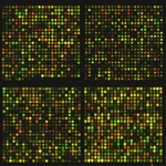
Applications in global gene expression analysis: Microarray and RNA sequencing
Global gene expression analysis, which involves studying the expression levels of all genes in a genome, can be performed using two main technologies: microarray and RNA sequencing (RNA-seq). Here’s an overview of how these technologies are used in global gene expression analysis:
- Microarray Technology:
- Principle: Microarrays consist of small glass slides or chips with thousands of DNA or RNA probes immobilized on their surface. These probes are complementary to specific genes or transcripts of interest. The sample RNA is labeled with a fluorescent dye and hybridized to the microarray. The intensity of the fluorescent signal at each probe spot indicates the expression level of the corresponding gene.
- Applications: Microarrays are used to study gene expression patterns in different conditions or tissues, identify biomarkers for disease diagnosis or prognosis, and discover novel genes or pathways involved in biological processes. They can also be used to study alternative splicing and post-transcriptional modifications.
- RNA Sequencing (RNA-seq):
- Principle: RNA-seq involves sequencing the entire transcriptome of a sample using high-throughput sequencing technologies. The RNA is converted to complementary DNA (cDNA), which is then sequenced. The resulting sequence reads are mapped to the reference genome or transcriptome to quantify gene expression levels.
- Applications: RNA-seq provides a comprehensive view of gene expression and allows for the identification of novel transcripts, isoforms, and non-coding RNAs. It is used to study gene expression changes in response to different conditions, identify disease-associated genes, and characterize transcriptional regulatory networks.
Comparison:
- Sensitivity and Dynamic Range: RNA-seq has a higher sensitivity and dynamic range compared to microarrays, allowing for the detection of low-abundance transcripts and more accurate quantification of gene expression levels.
- Cost and Throughput: Microarrays are generally more cost-effective for large-scale studies, while RNA-seq is more suitable for in-depth analysis of transcriptomes.
- Data Interpretation: RNA-seq provides more detailed information on transcript structure and alternative splicing compared to microarrays, but requires more computational resources for data analysis.
Overall, both microarray and RNA-seq technologies have their advantages and limitations, and the choice of technology depends on the specific research goals and experimental design.
Applications of recombinant DNA technology in medicine, agriculture, and veterinary science
Recombinant DNA technology, which involves the manipulation of DNA to create new combinations of genes, has a wide range of applications in medicine, agriculture, and veterinary science. Here are some key applications:
- Medicine:
- Production of Therapeutic Proteins: Recombinant DNA technology is used to produce therapeutic proteins, such as insulin, growth factors, and antibodies, which are used to treat various diseases, including diabetes, cancer, and autoimmune disorders.
- Gene Therapy: Recombinant DNA technology is used in gene therapy to treat genetic disorders by introducing functional genes into patients’ cells to correct genetic defects.
- Vaccine Development: Recombinant DNA technology is used to produce vaccines against infectious diseases, such as hepatitis B, HPV, and COVID-19, by expressing viral antigens in host cells to stimulate an immune response.
- Agriculture:
- Genetically Modified (GM) Crops: Recombinant DNA technology is used to create GM crops with improved traits, such as pest resistance, herbicide tolerance, and enhanced nutritional content. These crops can help increase yield, reduce pesticide use, and improve food security.
- Livestock Improvement: Recombinant DNA technology is used to improve livestock by introducing genes for desirable traits, such as disease resistance, improved growth rate, and enhanced milk production.
- Veterinary Science:
- Animal Health: Recombinant DNA technology is used to develop vaccines and treatments for animal diseases, such as foot-and-mouth disease, rabies, and bovine leukemia.
- Biomedical Research: Recombinant DNA technology is used in veterinary research to study animal genetics, disease mechanisms, and drug development.
- Environmental Applications:
- Bioremediation: Recombinant DNA technology is used to develop microorganisms with enhanced bioremediation capabilities, which can be used to clean up environmental pollutants, such as oil spills and toxic chemicals.
Overall, recombinant DNA technology has revolutionized the fields of medicine, agriculture, and veterinary science, leading to the development of new treatments, improved crop varieties, and enhanced animal health. However, it also raises ethical, environmental, and regulatory challenges that need to be addressed to ensure its responsible use and beneficial impact.
Related posts:
![humangenome]()
Step-by-Step Guide: Best Pipeline for Human Whole Exome Sequencing (WES)
bioinformatics![microarray analysis]()
Step-by-Step Guide: Analyzing Microarray Data in Bioconductor
bioinformatics![data-science]()
Data Science for Beginners: A Comprehensive Guide with a Focus on Bioinformatics
bioinformatics![bioinformatics software]()
2023's Comprehensive Guide to Choosing the Best Bioinformatics Software
bioinformatics![DNA-crispr]()
Step-by-Step Manual: Merging Many Small BAM Files into One Large BAM File
bioinformatics![R-perl-python-bioinformatics]()
Integrating NCBI Data Retrieval and Bioinformatics Analysis in Python, Perl, and R
Guides![DNA-crispr]()
Step-by-Step Guide: How to Draw a Heat Map for Gene Expression Data
bioinformatics![bioinformatics-internship]()
How to Secure a Bioinformatics Internship: Tips and Tricks
bioinformatics![Bioinformatics]()
How to Use Bioinformatics Databases and Tools to Accelerate Your Research
bioinformatics![python-bioinformatics-basics]()
Python vs. R for Genomic Data Analysis: A Comprehensive Guide
bioinformatics![will bioinformatics replace by AI]()
Deep Learning Models for Advanced 3D Protein Structure Prediction
bioinformatics![mesothelioma-cancer-bioinformatics]()
Navigating the Omics Landscape of Mesothelioma: A Comprehensive Guide
bioinformatics![insurance-bioinformatics]()
Revolutionizing Insurance with Bioinformatics, Genomics, and Omics: A Comprehensive Guide
bioinformatics![Neuroproteomics]()
Protein Extraction and Purification in Proteomics
Guides![Orf]()
Difference Between CDS and ORF: A Beginner’s Guide to Bioinformatics
bioinformatics![Genome analysis tools]()
A Beginner’s Guide to Visualizing Genomic Feature Data
bioinformatics